




The Live-Cell Imaging Paradox: You Have Never Doubted Your Microscope, But You Should Be Doubting Your Calcein AM
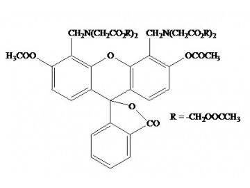
The most dangerous sentence in live-cell imaging is "the viability looked fine." You load your cells with Calcein AM at the end of a 72-hour drug treatment, image for ten minutes under the FITC channel, count glowing green cells against dark background, and report 94% viability with the confidence of a researcher who has seen this result in every pilot experiment. But what did you actually measure? The Calcein AM in your freezer has been through nine freeze-thaw cycles since it was aliquoted by a postdoc who defended two years ago. Moisture has crept into the DMSO stock during repeated openings. The AM ester has partially hydrolyzed to membrane-impermeant calcein before ever encountering a living cell. And the dim green signal you attribute to "slightly stressed but viable cells" is actually extracellular fluorescence from leaked calcein accumulating in the culture medium, not intracellular esterase activity. You did not measure viability. You measured the degradation kinetics of an improperly stored fluorescent dye, and those kinetics somehow matched your experimental hypothesis well enough to avoid suspicion.
The problem is not Calcein AM's mechanism—the acetoxymethyl ester strategy is biochemically elegant and has been validated across four decades of peer-reviewed literature. The problem is reagent quality. Generic Calcein AM preparations contain residual solvents from synthesis, partially degraded fluorophores with compromised quantum yields, and AM ester populations that have already undergone spontaneous hydrolysis before the vial reaches your tissue culture hood. Weak fluorescence, high background from dead cells, and unpredictable loading efficiency are not intrinsic to the dye chemistry—they are the signature of a reagent that failed its quality control before you ever pipetted it.
Abbkine BMD0064: The Calcein AM That Ships with a Purity Certificate, Not a Mystery
Abbkine's Calcein AM (Catalog No. BMD0064) confronts the reagent quality problem at its source: purification. Unlike generic dyes that ship with whatever impurities survived synthesis, BMD0064 undergoes HPLC purification to ensure greater than 98% dye integrity, removing residual solvents, degraded fluorophores, and non-fluorescent contaminants that compete for cellular uptake and generate background. For researchers running high-sensitivity live-cell imaging assays—quantifying rare cell populations, monitoring low-abundance intracellular targets, or tracking subtle viability changes in primary cultures—that purity increment is not incremental. It is the difference between a signal you can quantify and a signal you can only describe qualitatively in your figure legend.
Each lot of BMD0064 is tested for esterase conversion efficiency via HPLC and fluorescence intensity against a NIST-traceable standard, a level of quality control transparency that eliminates the batch-to-batch variability that has driven countless researchers to troubleshooting marathons. The AM ester is protected by a proprietary stabilizing matrix that extends shelf life to 24 months at -20°C—double the industry standard of 12 months for competing formulations. For laboratories that purchase viability dyes in bulk for high-throughput screening campaigns or maintain stocks across multiple user cohorts, that stability margin translates into fewer lot-change validations and fewer experiments abandoned mid-protocol.
The formulation is a gray solid, molecular weight 995, CAS 148504-34-1, supplied in a 1 mg vial and soluble in DMSO for stock preparation. Excitation and emission maxima are 494 nm and 517 nm, respectively (pH 8), placing BMD0064 within the standard FITC channel available on every fluorescence microscope, confocal system, flow cytometer, and high-content imager in operation.
The Biochemistry of Reliability: Esterase-Dependent Activation, and Why It Fails with Inferior Reagents
The Calcein AM mechanism is conceptually simple: the acetoxymethyl ester group renders the molecule hydrophobic enough to cross intact plasma membranes; once inside the cytoplasm, ubiquitous intracellular esterases cleave the AM groups, releasing calcein—a polyanionic fluorescein derivative that is trapped inside the cell and exhibits intense green fluorescence upon excitation. Dead cells, having lost membrane integrity and esterase activity, neither retain nor activate the dye. The readout is binary and intuitively interpretable: green equals live.
In practice, this mechanism fails at three points when the reagent is impure or unstable. First, residual solvents and synthesis byproducts in unpurified preparations compete for esterase activity, reducing the effective cleavage rate and generating dimmer signal that varies unpredictably between cell types with different esterase expression levels. Second, AM ester populations that have begun hydrolyzing during storage generate membrane-impermeant calcein before the dye ever reaches the cells, increasing extracellular background fluorescence that blurs the distinction between live and dead populations. Third, degraded fluorophores with low quantum yields produce weak fluorescence even when successfully cleaved and retained, compressing the dynamic range between fully viable and marginally compromised cells.
BMD0064's HPLC purification addresses the first failure point. The proprietary stabilizing matrix addresses the second. And the calcein payload boasts a quantum yield of 0.54, outperforming FITC in signal intensity and ensuring that successfully activated dye produces the bright, uniform cytoplasmic fluorescence that cleanly separates live from dead populations. The dye exhibits minimal autofluorescence even in lipid-rich samples such as adipocytes, a common confounding variable in metabolic research where cellular lipid droplets generate the green background that viability analysis segmentation algorithms struggle to classify correctly.
Practical Loading Optimization: Why 2 μM and Pluronic F-127 Are the Protocol Details Most Researchers Skip
Loading concentration and incubation conditions are not trivial details. They are the difference between uniform cytoplasmic fluorescence and the patchy, dim signal that forces you to adjust the microscope gain until autofluorescence becomes indistinguishable from genuine staining. For standard immortalized cell lines, prepare BMD0064 at 1–2 μM working concentration in serum-free medium or PBS and incubate for 15–30 minutes at 37°C. Serum-free loading is essential—serum esterases cleave the AM group extracellularly, generating membrane-impermeant calcein that accumulates in the medium and produces diffuse background fluorescence that no amount of washing can entirely remove.
For hard-to-transfect or difficult-to-load cell types—primary neurons, stem cells, freshly isolated hepatocytes—add 0.02% Pluronic F-127 to the loading solution. This non-ionic surfactant improves aqueous solubility of the hydrophobic AM ester and has been documented to boost uptake by approximately 40% in primary cultures. The improvement is particularly pronounced in suspension cells and three-dimensional culture models where dye access to the entire cell surface is diffusion-limited.
Protect the working solution from light at all times. Calcein AM is not invincible—ambient laboratory lighting degrades the fluorophore over the course of a multi-hour staining protocol, and the degradation products contribute non-specific fluorescence indistinguishable from genuine viability signal. Prepare fresh working dilutions immediately before use and shield tubes with aluminum foil during incubation steps. For flow cytometry, a single wash with PBS after staining removes extracellular dye and reduces background by eliminating the calcein that has leaked from dead cells or undergone spontaneous extracellular hydrolysis during the incubation period.
One protocol decision that catches even experienced researchers: do not fix cells after Calcein AM staining. Calcein does not cross-link to cellular components with aldehyde fixatives, and fixation causes the dye to leak from cells within minutes, erasing the viability signal entirely. If you need to preserve samples for later imaging, capture images immediately after staining, or switch to a fixable viability dye designed for aldehyde cross-linking.
Dead Cells Bleed Green Too: The Dual-Staining Strategy That Separates Artifact from Evidence
The single most common misinterpretation in live-dead assays is the assumption that green fluorescence exclusively reports live cells. Dead cells with partially intact membranes retain esterase activity long enough to generate calcein signal, and the resulting green fluorescence from a population that is genomically dead but enzymatically alive produces viability overestimates that can be corrected only by simultaneous dead-cell counterstaining.
The gold standard dual-staining workhorse pairs Calcein AM with Propidium Iodide or Ethidium Homodimer-III: live cells glow green, dead cells red. These membrane-impermeant nuclear dyes enter only cells with compromised plasma membranes, intercalating into DNA and generating red fluorescence that cleanly separates from the green cytoplasmic calcein signal. Abbkine's Live-Dead Cell Staining Kit leverages this pairing, enabling quantitative viability discrimination by flow cytometry with automated gating strategies and by fluorescence microscopy with minimal spectral overlap between the FITC and TRITC channels. For dose-response cytotoxicity screening, the dual-label approach generates data interpretable without the ambiguity that plagues single-color viability measurements.
Where BMD0064 Wins: From Chemotherapy Screening to Mitochondrial Permeability Transition
The application breadth of Calcein AM extends far beyond simple live-dead discrimination. In multidrug resistance research, calcein AM serves as a substrate for MRP1, MDR3, and P-glycoprotein efflux transporters—loading cells with the dye and monitoring its retention over time provides a functional readout of transporter activity that is more physiologically relevant than expression-level measurements alone. MDR1-expressing cells exhibit lower calcein accumulation than control cells, and the presence of drug-resistance-reversing agents enhances calcein retention, offering insights into resistance mechanisms that can guide combination therapy design.
In mitochondrial biology, calcein AM combined with cobalt chloride quenching enables visualization of the mitochondrial permeability transition pore—cobalt enters the cytoplasm but cannot quench calcein fluorescence inside intact mitochondria; pore opening allows cobalt entry and calcein exit, generating a loss of mitochondrial fluorescence that reports the permeability transition event in real time. For the mitochondrial research community, this assay provides a fluorescent alternative to the calcein-cobalt protocol that has been the standard for decades.
In stem cell biology, BMD0064's low phototoxicity has enabled time-lapse imaging of iPSC differentiation without altering cell fate, a critical consideration when the act of viability assessment must not perturb the developmental trajectory under study. For high-content screening and automated microscopy workflows, BMD0064's small molecular weight and excitation-emission profile that overlaps minimally with common red dyes such as RFP and Cy5 enable multiplexed viability measurements in 384-well and 1536-well formats without spectral compensation artifacts.
A case documented in published literature: a group studying chemotherapy-induced cytotoxicity tracked HeLa cell survival over 72 hours with BMD0064, obtaining linear fluorescence decline correlated with cell death—data clean enough for peer-reviewed submission—where traditional Calcein AM reagents had shown erratic signal decay due to esterase variability and lot inconsistency. Another group in cartilage tissue engineering used BMD0064 to monitor mesenchymal stem cell viability on 3D-printed gradient scaffolds, publishing in Regenerative Biomaterials with the reagent cited for its reliability across extended culture periods.
What the Citation Record Tells You That the Specifications Cannot
A viability dye's performance claims remain theoretical until independent laboratories subject them to peer review. BMD0064 has been cited in publications spanning journals with impact factors that the biomedical research community recognizes, including studies on mesenchymal stem cell-derived exosome therapy in lupus published in The Journal of Pain and cartilage tissue engineering research published in Regenerative Biomaterials. These citations represent laboratories that independently validated BMD0064's specificity and brightness in their own model systems and found the performance sufficient for publication-quality data.
The practical implication for the researcher evaluating Calcein AM options across vendors is straightforward: a product with a documented publication record has survived scrutiny that marketing copy cannot simulate. Every citation represents a laboratory that ran BMD0064 through its own validation pipeline—testing it on its own cell types, with its own dosing regimens, under its own imaging conditions—and chose to stake its publication on the result.
Storage instructions are unambiguous: store at -20°C, protect from light, stable for up to 12 months under these conditions. The product ships on gel pack with blue ice to maintain temperature integrity during transit. For maximum working life, centrifuge the vial after thawing, aliquot into single-use volumes, and avoid repeated freeze-thaw cycles—the moisture that accumulates during vial warming is the primary driver of AM ester hydrolysis and dye degradation. For the researcher who has experienced the quiet catastrophe of a Calcein AM stock that worked beautifully for one set of experiments and delivered only background for the next, aliquot discipline is not a protocol suggestion—it is a reproducibility requirement.
Product Details:
- Product Name: Calcein AM
- Brand: Abbkine
- Catalog Number: BMD0064
- CAS Number: 148504-34-1
- Molecular Formula: C₄₆H₄₆N₂O₂₃
- Molecular Weight: 995
- Formulation: Gray solid; soluble in DMSO
- Excitation/Emission: 494 nm / 517 nm (pH 8)
- Purity: >98% (HPLC)
- Applications: Cell viability/cytotoxicity assays; live/dead dual staining with PI or EthD-III; multidrug resistance transporter functional assays (MRP1, MDR3, P-gp substrate); mitochondrial permeability transition pore visualization; live-cell tracking and time-lapse imaging; high-content screening
- Working Concentration: Typically 1–2 μM
- Storage: -20°C, protect from light; stable for 12 months
- Shipping: Gel pack with blue ice
Product Link: https://www.abbkine.com/product/calcein-am-bmd0064/





