




Your Dead-Cell Stain Has Been Betraying You Since the Day You Thawed It. Abbkine's PI (BMD0065) Is the Only Propidium Iodide That Tells the Truth About Membrane Integrity.
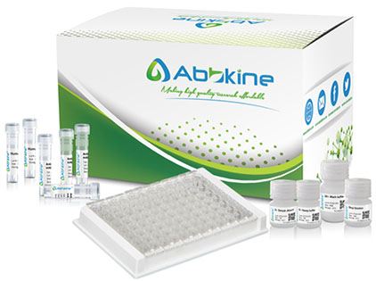
You have been there. The flow cytometer plots two populations where your eye insists there should be three: a tight cluster of live cells, a smear of debris, and a mysterious intermediate cloud that refuses to resolve into the clean early-apoptotic versus late-apoptotic gate your protocol promised. You increase the PI voltage, hoping sharper dead-cell discrimination will emerge, and instead the live-cell peak shifts rightward—every cell in your sample now fluoresces red, and you can no longer distinguish compromised membranes from intact ones. The problem is not your gating strategy, your apoptosis induction, or your compensation matrix. The problem is the propidium iodide in your freezer, a reagent you inherited from a predecessor who inherited it from a predecessor, its purity eroded by freeze-thaw cycles and its membrane impermeability compromised by degradation products that leak across intact plasma membranes and stain nucleic acids inside cells that are genomically alive.
Propidium iodide is defined by what it cannot do: it cannot cross an intact plasma membrane. This single biophysical constraint—the cationic charge that repels phospholipid head groups, the molecular weight of 668.4 that exceeds passive diffusion thresholds—is what makes PI the universal dead-cell marker in flow cytometry, fluorescence microscopy, and high-content screening. But that constraint is only as reliable as the reagent's purity. Low-grade PI preparations accumulate de-iodinated byproducts and positively charged degradation fragments that partition into live cells, generating the false-positive dead-cell signal that inflates apoptosis rates, distorts dose-response curves, and quietly invalidates cytotoxicity data that passes peer review only because reviewers cannot see what happened before the graph was generated.
Abbkine's Propidium Iodide (Catalog No. BMD0065) confronts this problem at the molecular level. With a molecular formula of C₂₇H₃₄I₂N₄, the product maintains consistent chemical purity validated via HPLC, ensuring that membrane impermeability remains absolute. The dye intercalates between base pairs of double-stranded DNA with high affinity, and upon binding, its fluorescence is enhanced 20- to 30-fold—the excitation maximum shifts to approximately 535 nm and the emission maximum to approximately 617 nm, producing the bright red signal that cleanly separates dead cells from live populations. The product's strict adherence to the defining characteristic of PI—selective membrane permeability—ensures it cannot penetrate the intact membranes of viable cells, guaranteeing exclusive staining of dead cells and late apoptotic cells where membrane integrity is compromised. Unlike low-quality PI alternatives that leak into live cells causing false positives, BMD0065 delivers the precision that apoptosis assays, cytotoxicity screens, and cell cycle analyses demand when the biological difference between 5% and 15% dead cells determines whether a drug candidate advances or dies.
The Three-Species Compatibility That Eliminates the Need for a Separate Dead-Cell Dye for Every Model System on Your Bench
What distinguishes BMD0065 from niche PI formulations is its broad cross-species compatibility, covering mammals, bacteria, and yeast—three of the most widely used model systems in biological research. This versatility aligns with the growing trend of cross-system validation: researchers can now use a single PI reagent to assess cell viability in mammalian cancer cell lines, bacterial cultures (e.g., E. coli), and yeast (e.g., Saccharomyces cerevisiae) without sacrificing specificity. In antibiotic susceptibility testing or antifungal drug screening, BMD0065's ability to stain non-viable microbial cells eliminates the need for separate dyes, streamlining workflows and reducing experimental variability. In mammalian research, it performs reliably across cell types—from adherent fibroblasts to suspension lymphocytes—making it a flexible tool for diverse applications, including cell toxicity assays, apoptosis studies, and cell cycle analysis.
The suggested working concentration ranges from 1–5 μM, a practical window that accommodates the staining intensity requirements of flow cytometry (where higher PI concentrations ensure stoichiometric DNA binding for cell cycle histogram resolution), fluorescence microscopy (where 1–2 μM typically suffices for clear dead-cell identification without detector saturation), and high-throughput screening formats (where consistent staining across hundreds of wells demands a concentration that balances signal strength with minimal well-to-well variability). The product is supplied as a ready-to-use liquid formulation, eliminating the reconstitution errors and DMSO exposure variability that plague powdered PI stocks and introduce an additional source of experiment-to-experiment inconsistency.
The Calcein AM-PI Axis: Why Your Viability Data Is Only as Strong as Your Dead-Cell Counterstain
Propidium iodide is rarely used alone, and its most powerful deployment is alongside Calcein AM in the live-dead dual-staining format that has become the gold standard for viability assessment across cell biology. Calcein AM crosses intact membranes and is cleaved by intracellular esterases into green-fluorescent calcein, retained exclusively in live cells. PI enters only cells with compromised membranes, intercalating into DNA and producing red fluorescence. Together, they generate the binary green-live, red-dead readout that is immediately interpretable by flow cytometry and fluorescence microscopy alike.
But the binary logic of this assay collapses when the PI component is impure. If degraded PI leaks into live cells, the green and red signals overlap, producing double-positive events that are neither live nor dead—they are artifacts of reagent failure, misclassified by automated gating algorithms as "apoptotic" or "compromised" and reported in figures that no reviewer can audit for dye quality. BMD0065's rigorous quality control—including HPLC purity verification, sterility testing, endotoxin limit checks below 0.1 EU/mg, and fluorescence intensity validation—ensures that the red channel reports only dead cells, preserving the assay's binary logic and generating data that withstand the scrutiny of gating reviewers and replication attempts alike.
For apoptosis detection, PI pairs with Annexin V conjugates to discriminate early apoptotic cells (Annexin V-positive, PI-negative, with intact membranes) from late apoptotic and necrotic cells (Annexin V-positive, PI-positive, with compromised membranes). Here again, PI that leaks into early apoptotic cells collapses the critical quadrant separation that defines the assay's interpretive framework. BMD0065's membrane impermeability, validated by HPLC purity, maintains this separation and ensures that early apoptosis rates are not inflated by dye leakage artifacts.
Practical Protocol Wisdom That Separates Publication-Grade Dead-Cell Discrimination from the Images Your Committee Will Question
The instructions packed with BMD0065 reflect the accumulated experience of troubleshooting PI staining across diverse cell types and detection platforms. The mandate is clear: centrifuge the product to the bottom of the tube before use to recover material that may have settled during storage, then proceed with subsequent experiments. Protect the working solution from light at all times—fluorescent dyes all face the problem of quenching, and minimizing light exposure and observing immediately slows the quenching. PI is generally considered carcinogenic; wear a lab coat and disposable gloves for safety.
For flow cytometry, resuspend stained cells in PBS and analyze immediately. PI fluorescence is collected in the FL2 or PE channel; do not fix cells after PI staining unless your protocol specifically validates fixative compatibility, as fixation can equalize membrane permeability and erase the live-dead distinction the assay was designed to detect. For fluorescence microscopy, a 30-minute incubation with PI at room temperature protected from light, followed by a gentle PBS wash, produces bright nuclear staining in dead cells with minimal background—extended incubation beyond 45 minutes risks PI redistribution and increased cytoplasmic background from slow membrane penetration into compromised-but-not-yet-dead cells.
For multiplexed experiments, BMD0065's fluorescence emission profile (~617 nm) is spectrally compatible with green fluorophores such as FITC, GFP, and Calcein AM as well as blue dyes like DAPI and Hoechst 33342, minimizing spectral overlap and simplifying multicolor panel design. The product can also be combined with 488 nm-excited fluorophores such as FITC and PE, enabling its integration into established flow cytometry panels without channel reconfiguration.
Storage: protect from light and store at -20°C, where the product remains stable for 24 months. Alternatively, storage at 4°C protected from light maintains stability for 12 months. The product ships on blue ice to maintain temperature integrity during transit. For maximum working life, aliquot into single-use volumes upon receipt to avoid repeated freeze-thaw cycles that degrade dye integrity and compromise membrane impermeability.
The $29 Question: Why Premium Performance Does Not Require a Premium Price Tag
Cost is not an abstract concern when your experimental design involves eighteen treatment groups, quadruplicate wells, and repeated viability measurements at six time points over a 72-hour cytotoxicity study. PI consumption scales linearly with sample number, and pricing that seems negligible for a single 12-well plate becomes a line item that competes with primary antibodies when the experiment scales to high-throughput dimensions.
BMD0065 is priced at 50 for the same quantity while maintaining comparable purity and performance metrics validated by HPLC and fluorescence intensity testing. For academic laboratories running large-scale viability screens and for core facilities servicing multiple research groups with standardized staining protocols, this cost differential compounds across the hundreds of staining reactions that a 5 mg supply can support. The product's long-term stability—24 months at -20°C stored in dark conditions—further reduces waste from degraded reagents, ensuring that the vial you open today performs identically to the vial you open eighteen months from now.
BMD0065 at the Intersection of Reproductive Toxicology, Antimicrobial Screening, and High-Throughput Drug Discovery
Industry trends in cell analysis increasingly favor reagents that balance performance, affordability, and ease of use—attributes that BMD0065 delivers across diverse research contexts. The rise of high-throughput screening in drug discovery demands dyes that work seamlessly with automated platforms; BMD0065's stable fluorescence, resistant to photobleaching during extended imaging sessions, and compatibility with standard assay buffers such as PBS and HBSS make it suitable for high-throughput workflows.
In reproductive toxicology—the context that drives demand for validated rat model reagents including the Abbkine Rat FSH and Rat GSH ELISA kits—PI serves as the terminal viability readout for granulosa cell cytotoxicity assays, Sertoli cell toxicant screens, and embryo culture quality control. In antimicrobial susceptibility testing, BMD0065's cross-species compatibility across bacteria and yeast eliminates the need to stock separate viability dyes for prokaryotic and eukaryotic targets, streamlining the comparative experimental designs that characterize modern antibiotic discovery pipelines. As a CAS 25535-16-4 compliant reagent, BMD0065 meets international quality standards, ensuring its compatibility with global research workflows and publication requirements.
The product has been cited in a growing body of peer-reviewed publications, representing laboratories that independently validated BMD0065's specificity and performance in their own model systems—from cancer biology to microbiology—and found the data sufficient for publication. Every citation represents a vote of confidence from a research group that ran the dye through its own validation pipeline and staked its results on the outcome.
Product Details:
- Product Name: Propidium Iodide (PI)
- Brand: Abbkine
- Catalog Number: BMD0065
- CAS Number: 25535-16-4
- Molecular Formula: C₂₇H₃₄I₂N₄
- Molecular Weight: 668.4
- Formulation: Ready-to-use liquid
- Excitation/Emission (DNA-bound): ~535 nm / ~617 nm
- Purity: Validated by HPLC
- Applications: Dead-cell staining; live/dead dual staining with Calcein AM; apoptosis detection with Annexin V; cell cycle analysis; bacterial and yeast viability assessment
- Working Concentration: 1–5 μM
- Storage: -20°C, protect from light, stable for 24 months; alternatively 4°C for 12 months
- Shipping: Gel pack with blue ice
Product Link: https://www.abbkine.com/product/pi-bmd0065/





