


The Near-Infrared Imperative: Why Your Visible-Spectrum Secondary Antibody Is Costing You More Than Signal
Biologists reach for far-red fluorophores like astronomers build infrared telescopes: longer wavelengths mean less interference from the medium. Tissue autofluoresces most aggressively in the blue-green range, where flavins, NADH, and lipofuscin bury weak antigen signals beneath a haze of false photons. Shift detection above 680 nm, and autofluorescence drops by orders of magnitude. Yet many labs default to green and red secondaries out of habit, accepting the 10:1 signal-to-noise ceiling on thick sections, then spending hours subtracting background that should never have been collected. The near-infrared channel itself has historically frustrated with dye aggregation, hydrolytic degradation, and emission tails that bleed into the 800 nm detection window—problems not of concept but of fluorophore chemistry. Abbkine’s DyLight 680, Goat Anti-Rabbit IgG (Catalog No. A23720) addresses these challenges with advanced dye engineering and rigorous antibody purification to deliver high signal-to-noise across imaging and quantitative blotting.
A23720: The Conjugate Engineered for the Odyssey Era, Not the Arc Lamp Era
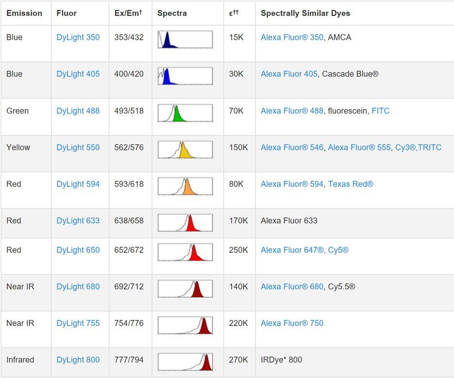
A23720 is a goat anti-rabbit IgG (H+L) near-infrared conjugate, affinity-purified on solid-phase rabbit IgG columns to greater than 95% purity and coupled to a sulfonate-modified cyanine fluorophore tuned precisely for the 700 nm channel (Ex/Em: 692/712 nm). These coordinates match the detection band of LI-COR Odyssey, Azure, and Bio-Rad fluorescence imagers, while maintaining sufficient separation from DyLight 800 and IRDye 800CW to enable clean two-color multiplexing without computational unmixing. The antibody recognizes heavy chains of rabbit IgG and light chains common to all rabbit immunoglobulin subclasses, with no reactivity against non-immunoglobulin serum proteins—broad subclass coverage that prevents differential staining artifacts when working with rabbit monoclonal antibodies of varying IgG types. Extensive cross-reactivity testing against mouse, human, and other species confirms specificity, ensuring the signal originates from your rabbit primary, not endogenous immunoglobulins. Supplied as a liquid in PBS (pH 7.4) with sodium azide, BSA, and 50% glycerol, the ready-to-use formulation eliminates lyophilization errors. Centrifuge after thawing and aliquot to avoid freeze-thaw degradation.
DyLight 680: The Fluorophore That Solves Solubility, Photostability, and Spectral Bleed Simultaneously
The DyLight 680 fluorophore employs sulfonate modification chemistry to simultaneously enhance water solubility—preventing dye aggregation that generates punctate background artifacts—and resist the hydrolytic degradation that erodes the quantum yield of legacy NIR dyes during storage. Monomeric conjugates eliminate the fluorescent specks routinely misinterpreted as genuine punctate staining by inexperienced microscopists. Photostability testing demonstrates that DyLight 680 outperforms Alexa Fluor 680 in bleaching resistance, making A23720 suitable for time-lapse imaging and quantitative fluorescence microscopy where consistent signal is essential. The dye remains stable from pH 4 to pH 9, maintaining performance across fixation, permeabilization, and mounting steps. Its negligible spectral bleed avoids crosstalk with visible-wavelength dyes, and in two-color NIR western blotting using LI-COR Odyssey systems, the 700/800 nm separation yields clean, independently quantifiable bands without mathematical unmixing.
A Practical Guide to Optimizing Assays with A23720: Dilution Ranges, Sample Preparation, and Application-Specific Strategies
For most fluorescent applications, start between 1:50 and 1:1,000; for LI-COR Odyssey and similar NIR imagers, a 1:10,000 dilution serves as a practical starting point, leveraging the sensitivity of laser-based detection. In flow cytometry, pre-incubate samples with 5% normal goat serum for 15 minutes at 4°C to block Fc receptors, then apply A23720 at 1:400—adjusting to 1:200 for low-affinity primaries or 1:600 for high-affinity ones. For western blotting, a 1:1,000 dilution with two-hour room-temperature incubation in standard blocking buffer has been validated in published studies; the stable NIR signal replaces chemiluminescent decay, enabling archivable, linear quantification. On thick tissue sections, PLP fixation preserves antigenicity better than formalin, and overnight incubation at 4°C ensures uniform penetration without fluorophore degradation. Protect working solutions from light, prepare fresh dilutions on ice, centrifuge the stock after thawing, and aliquot into single-use volumes.
The Autofluorescence Advantage: Why NIR Detection Wins in Brain, Liver, and Tumor Tissue
Brain sections loaded with lipofuscin, liver dense with flavin oxidases and porphyrins, tumor xenografts accumulating necrotic debris—all generate intense autofluorescence that peaks between 500 and 650 nm. At 700 nm and beyond, endogenous fluorophore extinction coefficients fall dramatically, translating directly into darker backgrounds and higher signal-to-noise ratios without post-acquisition processing. For multiplexed immunofluorescence requiring four to six antigens on a single section, the far-red channel fills a spectral niche that visible-wavelength fluorophores cannot occupy without unacceptable overlap penalties. A23720 adds the 700 nm channel to existing panels without demanding the spectral unmixing algorithms that become numerically unstable as fluorophore counts increase.
Ten Publications and a Commitment to Reproducibility: The Independent Validation That Counts
A23720 has been cited in 10 publications spanning Experimental Neurology, Journal of Hazardous Materials, International Journal of Molecular Medicine, and Journal of Virology—independent validations across N6-methyladenosine, PM2.5 toxicity, cardiac pathophysiology, and oncology research. Abbkine subjects each conjugate lot to molar saturation optimization, establishing the dye-to-antibody ratio that maximizes fluorescence while minimizing background, rather than applying a fixed conjugation chemistry. This lot-specific QC ensures the reproducibility that separates reliable commercial secondaries from those requiring re-titration with every shipment. Priced at approximately $40 per 100 μL, the conjugate fits large-scale rodent studies without exhausting antibody budgets. Pair A23720 with DyLight 800 for two-color NIR westerns, or combine with DyLight 488, 594, and other conjugates to build multi-channel panels across the visible-to-NIR spectrum with consistent buffer chemistry and storage requirements.
Product Details:
- Product Name: DyLight 680, Goat Anti-Rabbit IgG
- Brand: Abbkine
- Catalog Number: A23720
- Host: Goat
- Clonality: Polyclonal
- Immunogen: Rabbit IgG whole molecule
- Reactivity: Rabbit IgG (heavy chains and light chains); no reactivity against non-immunoglobulin serum proteins
- Conjugate: DyLight 680 (Ex/Em: 692 nm / 712 nm)
- Purification: Affinity purified on solid-phase Rabbit IgG (H&L); >95% purity by SDS-PAGE
- Applications: FCM, ICC, IF, WB; suggested starting dilution 1:50–1:1000 for fluorescent applications; 1:10,000 for LI-COR Odyssey
- Formulation: Liquid in PBS (pH 7.4), 0.02% sodium azide, 1% BSA, 50% glycerol
- Storage: Stable for one year at -20°C from date of shipment; ship on blue ice
Product Link: https://www.abbkine.com/product/dylight-680-goat-anti-rabbit-igg-a23720/


