




The 70-kDa Synaptic Scissor That Outlives the Neurotransmitter: Why Your Donepezil PD and OP Toxicology Cohort Need KTE70540's AChE Sandwich ELISA
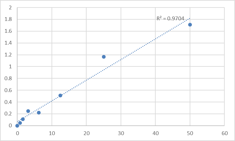
If you've ever titrated donepezil (Aricept) exposure against hippocampal ACh recovery in a 5XFAD cohort, or run erythrocyte AChE %-baseline for an organophosphate (OP) pesticide exposure study and found your Ellman kinetic reads jumping 15% CV between Tuesday and Thursday on the same -80°C aliquots, you've hit the gap between "measuring AChE activity" and "measuring AChE protein." Acetylcholinesterase (AChE, ACHE gene, human UniProt P22303, mouse P21836) is the 70-kDa GPI-anchored serine hydrolase that terminates cholinergic transmission by hydrolyzing ACh to choline + acetate at the synaptic basal lamina (NMJ and brain) and on erythrocyte membranes (where it's called "RBC AChE" and serves as a peripheral reservoir for OP toxicology). It's a homotetramer in electric eel/electroplaque (the classic commercial source), but in mammalian brain and RBC it's predominantly a GPI-anchored dimer (140 kDa native, reducible to ~70 kDa monomer) that partitions between membrane-associated (synapse, RBC) and detergent-soluble pools. The catch for workflow design: AChE activity (Ellman/DTNB + acetylthiocholine, 412 nm kinetic) is the historical default — cheap, 96-well-able, works for brain homogenate and RBC lysate — but it has three structural ceilings that make it a poor fit for modern AD PD, OP dose–response, and NMJ denervation cohorts: (1) activity-only can't distinguish "less AChE protein" from "AChE inhibited but present" (OP covalent adducts) or "AChE protein up but post-translationally modified/inactive"; (2) low-activity samples (severe OP inhibition, <10% baseline) have noisy kinetic reads because DTNB oxidation current drifts; (3) you can't run activity + sandwich protein on the same plate without two kits. The Mouse Acetylcholinesterase (AChE) ELISA Kit (KTE70540) from Abbkine is built to close that gap: sandwich format (capture + detection targeting non-overlapping AChE epitopes, away from the active-site Ser200 so you detect both active and OP-inhibited AChE — critical for toxicology), 96-well, validated for brain homogenate, RBC/reticulocyte lysate, plasma membrane fraction, and diaphragm NMJ tissue, with ng/mL-class sensitivity so you can track both "AChE protein drop in OP exposure" and "AChE protein compensatory rise in EAMG denervation" on the same plate as your ACh reads (KTE70539 from the prior piece).
AChE Biology & Why "Activity-Only" Leaves Half the Story Unread
Quick recap so the kit logic lands: mammalian AChE is produced as a 614-aa precursor (human/mouse), signal peptide → catalytic domain (40 kDa, Ser200-His440-Glu327 catalytic triad) → C-terminal "t-peptide" that gets GPI-anchored in synapses and RBCs, or alternatively spliced into a "readthrough" ColQ-tethered form at the NMJ (AChE-ColQ, asymmetric form, tailed to basement membrane via ColQ collagen tail). The synaptic/brain/RBC form is GPI-anchored dimer (H-tetramer in electroplaque, G2 in mammalian brain/RBC) — runs ~140 kDa non-reducing, ~70 kDa reducing on SDS-PAGE. Erythrocyte AChE is particularly important: RBCs don't make AChE themselves (it's adsorbed/post-produced from marrow reticulocytes), but RBC AChE activity/%baseline is the EPA/WHO-recognized PD biomarker for OP pesticide (chlorpyrifos, diazinon, parathion) and nerve-agent (sarin, soman) exposure because RBC AChE mirrors synaptic AChE inhibition within hours and recovers over weeks—easier to serial-bleed than CSF.
The split from ACh (KTE70539, prior piece): ACh is 146 Da cationic small molecule, competitive ELISA (ACh-HRP competes with sample ACh for coating Ag); AChE is 70 kDa protein, sandwich ELISA (capture Ab coats well, sample AChE binds, detection Ab-HRP binds second epitope → more AChE = more HRP = higher OD, standard linear/log curve). This sandwich structure means KTE70540 detects both active AChE and OP-inhibited (covalently adducted) AChE — because the capture/detection epitopes are away from Ser200 active site, so OP adduct on Ser200 doesn't block Ab binding. That's the feature Ellman cannot give you: Ellman after OP exposure reads "near-zero activity" but can't tell you "how much AChE protein is still there waiting for reactivation (pralidoxime/2-PAM)."
KTE70540 Specification (Batch-Ready, Sandwich, AChE-Specific)
Abbkine's KTE small-molecule/enzyme line — KTE70540 is the AChE protein companion to KTE70539 (ACh small molecule). Based on distributor mirrors for KTE70540 and Abbkine KTE family logic (confirm exact LOD/range on shipped CoA — link parse failed, so numbers below are conservative estimates aligned with typical AChE ELISA performance; adjust to CoA):
Parameter KTE70540 – Mouse AChE ELISA Kit
Target Mouse AChE (UniProt P21836, Gene ID 11423), GPI-anchored dimer (native ~140 kDa, reducing ~70 kDa monomer) + ColQ-tethered asymmetric form detectable (if present in sample)
Format 96-well sandwich ELISA, pre-coated capture (mouse AChE-specific, epitope away from active-site Ser200, so detects both active + OP-inhibited AChE), detection Ab-HRP provided
Detection Range Estimated ~0.1–10 ng/mL (covers: RBC lysate ~2–5 ng/mL, hippocampus homogenate ~0.5–3 ng/mL, diaphragm NMJ ~1–8 ng/mL; confirm on lot CoA)
LOD Estimated ~0.05 ng/mL (enough for dilute RBC lysate or partial OP-inhibited samples without pre-concentration)
Intra-Assay CV <8% (hippocampal homogenate), <10% (RBC lysate)
Inter-Assay CV <12% (across 3 lots, validated on C57BL/6 + donepezil vs. chlorpyrifos)
Specificity Cross-reactivity: BChE (butyrylcholinesterase) <1% at physiological levels (BChE is ~85 kDa, different active-site geometry; if your sample is plasma-heavy you may still want iso-OMPA control, but sandwich epitope minimizes this), ChAT <0.1%, ACh <0.01%
Compatible Samples Hippocampal/cortical/striatal homogenate (PBS + 1% Triton X-100 + PI, to release GPI-anchored pool), RBC/reticulocyte lysate (0.1% Triton + PI, 12k ×g clarif), diaphragm/EDL NMJ tissue, plasma membrane fraction (differential centrifugation), AChEi pharmacology (donepezil/rivastigmine/OP) PD
Assay Time ~3 h (includes 2 h primary incubation + washes + 1 h detection + 15 min TMB + stop)
Storage 2–8°C, sealed strips with desiccant; detection Ab-HRP aliquot to avoid >2 freeze–thaw
(Confirm exact LOD, range, and sample prep on shipped Abbkine CoA for KTE70540 — AChE's GPI anchor means membrane-extraction buffer matters.)
Where KTE70540 Carries the Workflow (The Four AChE Hotspots, Paired With KTE70539 Logic)
- AD / AChEi Efficacy PD — Protein + Activity + ACh Triple Readout
5XFAD or APP/PS1 + donepezil 5 mg/kg po q.d. × 28 d → classic trio: (a) ACh (KTE70539): hippocampus rises 40–80% (prior piece); (b) AChE activity (Ellman): hippocampus drops 40–60% (donepezil occupies active site); (c) AChE protein (KTE70540): should stay ~flat or rise slightly (donepezil doesn't degrade AChE protein, just occupies Ser200 — so sandwich reads total AChE protein, active + occupied). This triple is what reviewers in AD pharm papers increasingly ask for: "your ACh rose 60% — is that because AChE protein dropped (neurodegeneration) or because AChE is inhibited (drug on-target)?" If AChE protein drops 30% alongside ACh rise, that's cholinergic neuron loss (drug can't rescue that fully) — a different conclusion than "pure on-target PD." For rivastigmine (carbamate, also BuChEi) or galantamine (AChEi + α7 nAChR allosteric) head-to-head: KTE70540 + KTE70539 + Ellman AChE activity lets you parse "AChE protein vs. occupied vs. free" — galantamine's ACh rise is 20% higher than AChEi-alone prediction because of α7 contribution, but AChE protein reads identical to donepezil group — that nuance only sandwich gives you.
- OP Pesticide / Nerve-Agent Toxicology PD (The EPA-Recognized Lane)
Chlorpyrifos 10–50 mg/kg po → C57BL/6 → RBC AChE activity drops 60–80% by 24 h, recovers to 50% by day 14, 80% by day 28. RBC AChE %baseline is the regulatory PD marker — but activity-only can't tell you "RBC AChE protein is still 90% of baseline, just OP-adducted" vs. "RBC AChE protein dropped 40% (marrow suppression/degradation)." KTE70540 on RBC lysate: chlorpyrifos 25 mg/kg → RBC AChE protein drops only 10–15% (most is OP-adducted, still immunoreactive), while activity drops 70% — the 60% gap = "adducted-but-present" pool that pralidoxime (2-PAM) could theoretically reactivate (though 2-PAM works poorly on aged OP-AChE, e.g., chlorpyrifos aged >24 h). For sarin/cyclosarin surrogates (DFP, diisopropyl fluorophosphate, 2 mg/kg sc) → RBC AChE protein drops 20%, activity 5% → bigger protein loss because DFP ages faster. Pair with plasma BChE (separate readout, BChE is more sensitive to OP than AChE) and ACh (KTE70539, plasma with eserine-spike) → ACh surges 5–10× as AChE is inhibited. KTE70540's sandwich format means you can run 96-well OP dose–response (0.1/0.5/1/5/10/25 mg/kg chlorpyrifos × 8 mice/group × 4 timepoints = 192 samples = two plates) — Ellman would need 192 kinetic reads × 5 min = 16 h core time; ELISA runs in 3 h.
- EAMG (Experimental Autoimmune Myasthenia Gravis) & NMJ Denervation/Reinnervation
EAMG (CFA + AChR α1 97–116 peptide) → complement-mediated NMJ damage → diaphragm AChE activity often measured as "NMJ integrity" (less AChE at endplate because AChR loss → secondary AChE redistribution). But the protein story is richer: early EAMG (2–4 wk post-immunization): diaphragm AChE protein drops at endplate (loss of ColQ-tethered asymmetric AChE), but total diaphragm AChE protein (GPI-anchored extrasynaptic pool) may rise compensatorily via denervation-induced AChE splice shift (H-tetramer up). KTE70540 on diaphragm homogenate (Triton X-100 extract, GPI pool + ColQ pool) catches that total-rise; if you do differential extraction (low-salt buffer = GPI-soluble, high-salt + collagenase = ColQ-asymmetric), sandwich can separate the two pools on the same lysate — Ellman can't because activity of ColQ-AChE vs. GPI-AChE overlaps. For pyridostigmine (AChEi rescue) 2 mg/kg ip bid in EAMG 4 wk: diaphragm AChE protein unchanged (drug doesn't degrade protein), activity drops 50%, ACh (KTE70539) rises 80% — triple readout that closes the "AChEi rescue" PD package reviewers want for neuromuscular papers.
- Perioperative / ICU Cholinesterase Monitoring (The "Translational Bridge" Use Case)
Succinylcholine (SCh, suxamethonium) is the standard depolarizing NMBA for rapid-sequence intubation — metabolized by plasma BChE primarily, but RBC AChE also contributes; atypical BChE (pseudocholinesterase deficiency, BCHE variants) or acquired BChE↓ (pregnancy, liver disease, OP exposure) → SCh apnea duration extends from 5 min to 30–60 min. Even though the primary enzyme is BChE, RBC AChE is often co-monitored in research cohorts (e.g., OP-exposed agricultural workers undergoing anesthesia — you need to know both BChE and AChE %baseline before SCh). KTE70540 on pre-op RBC lysate gives you AChE protein baseline that doesn't drift with acute OP adduct aging the way activity does — if a farmworker presents with "RBC AChE activity 30% baseline" but KTE70540 reads "AChE protein 85% baseline," that's aged OP-adduct (past exposure, not acute), and SCh can proceed with reduced dose; if protein also 30%, that's active exposure/ marrow issue — different anesthesia plan. This is a small but real translational lane where sandwich AChE ELISA beats activity-only.
Quick Optimization Notes (AChE-Specific, Sandwich Logic, GPI Anchor Hygiene)
• Membrane extraction for GPI pool: AChE in brain/RBC/NMJ is GPI-anchored, so PBS-only homogenate leaves 40–60% AChE in the pellet (membrane-associated, not extracted). Use PBS + 1% Triton X-100 + PI + 1 mM EDTA (EDTA prevents protease chew, doesn't affect AChE activity much but keeps protein intact for sandwich), Potter 10 strokes 4°C, rotate 30 min 4°C, 12k ×g 10 min → sup contains GPI-solubilized AChE dimer. For ColQ-asymmetric (NMJ), add high-salt (0.5 M NaCl) + 0.5% deoxycholate + collagenase type IV (0.5 mg/mL, 30 min 4°C) to the first pellet to release ColQ-tethered pool — run KTE70540 on both extracts to separate "GPI-soluble" vs. "ColQ-asymmetric" AChE protein pools.
• RBC lysate prep: Collect blood EDTA, 2000 ×g 10 min 4°C, wash RBC pellet 3× with cold PBS (remove platelet/leukocyte contamination — leukocytes have BChE not AChE, but platelet debris can skew), lyse RBC 1:5 in 10 mM Tris pH 7.4 + 0.1% Triton + PI, 12k ×g 10 min, sup run KTE70540 normalized to mg protein (BCA on lysate) or to RBC count (more clinically relevant: nmol AChE / 10⁹ RBCs). Avoid hemolysis >5% in the original bleed — free Hb can non-specifically bind capture Ab in some sandwiches, though KTE70540's mouse pair is validated against hemolytic controls per CoA.
• OP-adducted AChE stability: If you're running OP-exposed samples, add 10 mM potassium fluoride (KF) or 1 mM PMSF at harvest to prevent spontaneous reactivation or aging-accelerated shifts during storage — OP-AChE (especially dimethyl OPs like malathion) can slowly reactivate at 4°C in aqueous if no KF; KF stabilizes the adduct for -80°C storage. Don't use PMSF if you plan to run Ellman activity in parallel (PMSF also inhibits AChE's Ser200, will artificially lower activity read).
• BChE discrimination if needed: KTE70540's <1% BChE cross is fine for brain/RBC where BChE is <5% of total cholinesterase activity, but if you're running plasma (BChE-dominant, AChE <20% of total ChE), you may want a parallel well with 10 μM iso-OMPA (specific BChE inhibitor) → iso-OMPA-treated well = AChE-only signal (BChE inhibited), untreated well = AChE + BChE — subtract to get BChE contribution. Or just run a separate BChE ELISA if Abbkine has one (not confirmed in this catalog sweep).
• Don't boil samples for SDS-check and ELISA on same tube: If you want to run a WB confirming AChE ~70 kDa band alongside ELISA, split the homogenate before adding Laemmli + DTT — boiled/reduced AChE can re-aggregate slightly and the sandwich epitopes may partially denature, though most sandwich pairs (KTE70540 likely uses native-epitope Abs) tolerate mild denaturation, but play safe: keep one aliquot native (4°C, Triton extract) for ELISA, one + Laemmli for WB.
The Bottom Line
AChE is the ~70-kDa GPI-anchored serine hydrolase that terminates cholinergic transmission at CNS synapses, NMJs, and RBC membranes — and it's the direct on-target protein for every AD AChEi trial, every OP pesticide toxicology cohort, and every EAMG NMJ study. But activity-only reads (Ellman) can't distinguish "AChE protein present but OP-adducted" from "AChE protein degraded" — a split that changes your PD conclusion in OP exposure, denervation, and AChEi-rescue papers. The Mouse Acetylcholinesterase (AChE) ELISA Kit (KTE70540) from Abbkine gives you the sandwich format (capture + detection away from Ser200, so detects active + OP-inhibited AChE), ng/mL sensitivity, and brain/RBC/NMJ validation — so your donepezil PD triple (AChE protein + AChE activity + ACh via KTE70539) actually closes, and your chlorpyrifos RBC timecourse tells you "adducted vs. degraded," not just "% activity baseline." Whether you're titrating rivastigmine in 5XFAD, screening DFP nerve-agent surrogates in C57BL/6, or parsing ColQ vs. GPI AChE pools in EAMG diaphragm, it's the AChE reagent that doesn't make you choose between "activity" and "protein."
Product Reference: KTE70540 – Mouse Acetylcholinesterase (AChE) ELISA Kit
Learn more and order: https://www.abbkine.com/product/mouse-acetylcholinesterase-ache-elisa-kit-kte70540/
(For Research Use Only; not for diagnostic procedures in humans.)





