


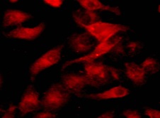
Visualize the Edge of Life: Advanced Orange Fluorescent Staining for Dynamic Plasma Membrane Studies
The plasma membrane defines the very boundary of cellular existence, a selectively permeable and astonishingly dynamic interface where critical signals are received, nutrients enter, waste products exit, and cellular identity is maintained. Accurately visualizing this thin, fluid bilayer in living cells is a cornerstone technique for countless discoveries in cell biology, immunology, and neurobiology. While green fluorescent probes are common, the need for multiplexing, compatibility with common green fluorescent protein (GFP) channels, or simply a preference for a longer wavelength signal often demands a reliable alternative. The TraKine™ Cell Plasma Membrane Staining Kit (Orange Fluorescence, KTC4002) from Abbkine meets this demand with a bright, photostable, and exceptionally membrane-selective orange fluorescent dye. Designed for minimal cellular disturbance, this kit provides a rapid and robust method to outline cell contours with high contrast, making it an ideal choice for live-cell imaging, co-localization studies with green-labeled targets, and flow cytometric analysis.
The Critical Need for Multi-Color Membrane Imaging in Complex Assays
Modern experimental designs frequently require the simultaneous observation of multiple cellular components. A researcher studying receptor internalization might need to track a GFP-tagged receptor while also visualizing the plasma membrane itself. In co-culture experiments, distinguishing the membranes of two different cell types is essential. Green fluorescence channels are often occupied by ubiquitous markers like GFP, FITC-conjugated antibodies, or viability dyes. The orange fluorescent probe (typically exhibiting excitation/emission maxima around ~549/576 nm, compatible with TRITC/RFP filter sets) in this kit fills a vital spectral niche. It allows for clear separation from blue (DAPI/Hoechst), green (FITC/GFP), and far-red signals, enabling sophisticated multi-parameter imaging and analysis without spectral bleed-through complications.
Superior Specificity Through Engineered Lipophilic Anchors
Unlike some general lipophilic dyes that rapidly internalize or stain intracellular membranes, the proprietary dye in the TraKine™ Orange kit is engineered for superior plasma membrane retention. Its molecular structure allows for fast, uniform insertion into the outer leaflet of the lipid bilayer. A key feature is its designed resistance to "flip-flop" across the membrane and to active transport mechanisms that would cause internalization. This results in a crisp, ring-like staining pattern that faithfully represents the true cell surface for extended periods, often lasting through typical live-cell imaging sessions. This stability is crucial for time-lapse studies of membrane dynamics, such as observing morphological changes during cell migration, division, or apoptosis.
Key Benefits That Streamline Research Workflows
The TraKine™ Cell Plasma Membrane Staining Kit (Orange Fluorescence, KTC4002) is optimized for performance and ease of use, offering distinct advantages:
• Excellent Photostability: The orange fluorophore exhibits reduced photobleaching compared to some green dyes, allowing for longer imaging sessions and the collection of more data points with consistent signal intensity.
• Low Cytotoxicity & Live-Cell Friendly: The staining protocol uses a dye concentration and formulation proven to be non-cytotoxic, ensuring that cell health, proliferation, and natural behaviors are not compromised during observation.
• Rapid, One-Step Staining: The process is remarkably simple. After a brief incubation of cells with the working dye solution (typically 5-15 minutes at 37°C or on ice), a gentle wash is all that's needed before imaging or analysis. No fixation, permeabilization, or complex steps are required.
• Broad Cellular Compatibility: The kit effectively stains a wide range of mammalian cell types, including both adherent and suspension cells, primary cells, and many delicate cell lines.
• Versatile Application Range: It is perfectly suited for a variety of techniques: confocal microscopy for high-resolution 3D membrane reconstruction, widefield fluorescence microscopy for routine checks and time-lapse, and flow cytometry for quantitative analysis of cell surface labeling in large populations.
• Ready-to-Use Components: The kit includes a concentrated dye solution and an optimized diluent/buffer, ensuring reproducible results from experiment to experiment.
A Simple Protocol for Immediate, High-Quality Results
Achieving clear plasma membrane staining is straightforward. Grow cells under standard conditions. Prepare the working stain by diluting the provided dye concentrate in the assay buffer or a serum-free medium. Replace the cell culture medium with this staining solution and incubate. For most applications at 37°C, 5-10 minutes is sufficient. For flow cytometry or experiments requiring minimized internalization, staining can be performed at 4°C for 15-20 minutes. After incubation, remove the staining solution and wash the cells once or twice with pre-warmed buffer or culture medium. The cells are now ready for immediate observation. The entire procedure, from staining to imaging, can often be completed in under 30 minutes.
Powerful Applications Across Diverse Research Fields
- Multiplex Live-Cell Imaging: Perfectly complement GFP/YFP-based reporters or stains. Clearly visualize the plasma membrane in orange while simultaneously tracking GFP-tagged organelles, cytoskeletal components, or signaling proteins in the green channel.
- Real-Time Analysis of Membrane Dynamics: Study rapid processes like phagocytosis, macropinocytosis, viral entry, or membrane blebbing with high temporal and spatial resolution, as the stable orange outline provides a constant reference for morphological changes.
- Cell-Cell Interaction and Synapse Studies: In immunology or neuroscience, precisely delineate the contact zones between immune cells and their targets or between neurons to analyze synapse formation, stability, and signaling.
- Membrane Integrity and Cytotoxicity Assays: Use the continuous orange ring as a baseline to quantify membrane damage. The disruption or loss of fluorescence uniformity is a direct and visual indicator of compromised membrane integrity caused by toxins, drugs, or physical stress.
- Flow Cytometry Gating and Normalization: Provide a bright, uniform fluorescent signal on the cell surface that can be used for accurate live-cell gating, distinguishing single cells from debris or aggregates, and normalizing the intensity of other fluorescent markers to cell size.
- Educational Tool for Cell Biology: The vivid, specific staining offers a striking visual demonstration of cell shape and membrane structure for teaching purposes in academic laboratories and classrooms.
Empowering Discovery with Clear Spectral Separation
In the colorful world of fluorescence microscopy and cytometry, having the right tool for clear spectral separation is not a luxury—it's a necessity for rigorous science. The TraKine™ Cell Plasma Membrane Staining Kit (Orange Fluorescence) provides researchers with a reliable, high-performance tool that excels in complex, multi-color experimental setups. By delivering a bright, specific, and stable signal in an underutilized spectral range, it solves common challenges in co-localization and multiplexing. This kit enables clearer insights into the dynamic life at the cell's edge, making it an essential reagent for any lab focused on cell surface biology, live-cell imaging, and advanced cytometric analysis.
Product Reference: KTC4002 – TraKine™ Cell Plasma Membrane Staining Kit (Orange Fluorescence)
Learn more and order: https://www.abbkine.com/product/trakine-cell-plasma-membrane-staining-kit-orange-fluorescence-ktc4002/


