


Mapping the Mitochondrial Network: Advanced Green Fluorescent Staining for Live-Cell Analysis and Beyond
Mitochondria are not just static bean-shaped organelles; they form a dynamic, interconnected network that constantly undergoes fusion and fission, processes intimately linked to cellular health, metabolism, and fate. Visualizing these intricate structures in their native, living state is paramount for research in cell biology, neurobiology, cancer metabolism, and toxicology. However, achieving specific, bright, and non-toxic staining of mitochondria has been a persistent challenge. The TraKine™ Mitochondrion Staining Kit (Green Fluorescence, KTC4003) from Abbkine provides a sophisticated solution. It utilizes a cell-permeant, cationic green fluorescent dye that selectively accumulates in active mitochondria, driven by the organelle's membrane potential. This kit delivers a robust, user-friendly method for high-contrast mitochondrial imaging in live cells, making it an indispensable tool for studying morphology, function, and dynamics.
The Central Role of Mitochondria and the Imperative for Precise Imaging
As the primary sites of ATP production through oxidative phosphorylation, mitochondria are fundamental to cellular energetics. Beyond power generation, they are critical hubs for calcium signaling, regulation of apoptosis (programmed cell death), and the biosynthesis of key metabolites. Their morphology—ranging from fragmented granules to elongated tubules—is a key indicator of their functional state and cellular health. Accurate visualization is therefore essential to investigate how mitochondrial dynamics correlate with metabolic shifts, stress responses, drug effects, and disease progression, such as in neurodegenerative disorders, cardiomyopathies, and cancer.
Mechanism of Action: Harnessing the Mitochondrial Membrane Potential
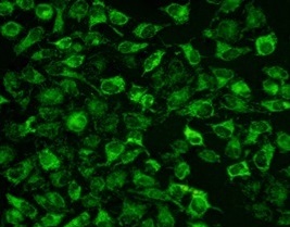
The core technology of this kit relies on a positively charged (cationic), lipophilic dye that readily crosses the plasma membrane. Once inside the cytosol, the dye is actively sequestered into the mitochondrial matrix. This accumulation is driven by the highly negative inner mitochondrial membrane potential (ΔΨm), a hallmark of healthy, respiring mitochondria. The dye binds to mitochondrial lipids or proteins, where its fluorescence is greatly enhanced, resulting in intense green fluorescence (with typical excitation/emission maxima around ~490/516 nm, compatible with standard FITC/GFP filter sets). Crucially, the staining intensity is proportional to the ΔΨm, allowing the signal to serve not only as a structural marker but also as a semi-quantitative indicator of mitochondrial health and activity.
Distinct Advantages Over Traditional Staining Methods
The TraKine™ Mitochondrion Staining Kit (KTC4003) offers significant improvements over conventional dyes like JC-1 or MitoTracker® analogues:
• High Specificity & Low Cytoplasmic Background: The dye exhibits exceptional selectivity for mitochondria over other cellular compartments, yielding crisp images with minimal off-target staining.
• Superior Photostability: The fluorophore is engineered for resistance to photobleaching, enabling longer time-lapse imaging sessions and the collection of more data points without significant signal loss.
• Live-Cell Compatibility & Low Toxicity: The optimized formulation allows for staining with minimal impact on mitochondrial function and overall cell viability, supporting long-term live-cell experiments.
• Rapid and Simple Protocol: Staining is typically complete within 15-30 minutes at 37°C. The protocol involves simply adding the diluted dye to cells, incubating, and optionally performing a gentle wash—no complex loading procedures or washes are required.
• Compatibility with Fixation: While ideal for live cells, the dye signal is often retained after fixation with paraformaldehyde, allowing for subsequent immunostaining or analysis at a later time.
• Broad Application Scope: The kit is effective for staining a wide variety of mammalian cell lines, primary cells, and even certain plant and yeast cells, making it a versatile choice for diverse research fields.
Streamlined Workflow for Reliable Results
The procedure is designed for simplicity and consistency. Cells are cultured under standard conditions. The provided concentrated dye is diluted in pre-warmed, serum-free culture medium or buffer to create a working solution. This solution is added directly to the cells and incubated in a standard cell culture incubator (37°C, 5% CO₂) for 15-30 minutes. For suspension cells, staining can be performed in centrifuge tubes. After incubation, the staining solution can be replaced with fresh, pre-warmed culture medium for immediate live-cell imaging. For some applications, a brief wash step may be included. The cells are then ready for observation using fluorescence microscopy, confocal microscopy, or analysis by flow cytometry or microplate readers.
Diverse Applications in Cutting-Edge Research
- Mitochondrial Morphology and Dynamics: Clearly visualize and quantify changes in mitochondrial network structure—fusion, fission, fragmentation, or elongation—in response to metabolic stimuli, pharmacological agents, or genetic manipulations.
- Assessment of Mitochondrial Health and Membrane Potential: Use the fluorescence intensity as a relative measure of ΔΨm. A decrease in signal intensity often indicates mitochondrial depolarization, a key early event in apoptosis or metabolic dysfunction.
- Co-localization Studies: Perfectly combine with other fluorescent probes (e.g., red fluorescent dyes for lysosomes, ER, or nuclei) to study organelle interactions, such as mitophagy or mitochondrial-ER contact sites.
- Cytotoxicity and Drug Screening: Efficiently screen compounds for mitochondrial toxicity by observing changes in staining pattern, intensity, or overall mitochondrial mass, which are common indicators of drug-induced injury.
- Apoptosis Detection: Monitor the early stages of apoptosis, characterized by mitochondrial membrane depolarization and fragmentation, often preceding other apoptotic markers like phosphatidylserine externalization.
- Stem Cell and Metabolic Research: Investigate metabolic shifts during stem cell differentiation or in cancer cells (Warburg effect) by correlating mitochondrial morphology and membrane potential with metabolic state.
- Neuroscience Research: Study mitochondrial trafficking and localization in neurons, which is critical for understanding synaptic function and neurodegenerative diseases.
An Essential Key to Unlocking Mitochondrial Function
Understanding the mitochondrion is understanding a core regulator of life, death, and disease. The TraKine™ Mitochondrion Staining Kit (Green Fluorescence) empowers researchers with a reliable, high-performance tool to illuminate these vital organelles with exceptional clarity and specificity. By combining ease of use with excellent performance in live cells, it removes technical barriers and provides a clear window into mitochondrial biology. This kit is a fundamental asset for any laboratory focused on cell metabolism, signal transduction, toxicology, or the pathophysiology of diseases where mitochondria play a central role.
Product Reference: KTC4003 – TraKine™ Mitochondrion Staining Kit (Green Fluorescence)
Learn more and order: https://www.abbkine.com/product/trakine-mitochondrion-staining-kit-green-fluorescence-ktc4003/


