


Illuminating the Cellular Frontier: A Guide to Precise Plasma Membrane Staining with Green Fluorescence
The plasma membrane is far more than a simple barrier; it is a dynamic, information-rich interface that governs every critical interaction between a cell and its environment. Visualizing this vital structure with clarity and specificity is a fundamental requirement in cell biology, enabling researchers to study cell morphology, membrane integrity, receptor localization, endocytosis, exocytosis, and cell-cell interactions. While numerous staining methods exist, many suffer from drawbacks such as poor specificity, slow kinetics, toxicity, or complex protocols that can perturb the very processes under observation. The TraKine™ Cell Plasma Membrane Staining Kit (Green Fluorescence, KTC4001) from Abbkine provides a rapid, highly selective, and exceptionally gentle solution for labeling the live cell plasma membrane. Utilizing a novel, non-cytotoxic green fluorescent probe, this kit delivers bright, stable staining within minutes, making it an indispensable tool for live-cell imaging, flow cytometry, and a wide array of cellular assays.
Why Precise Plasma Membrane Labeling is Non-Negotiable in Modern Research
Accurately distinguishing the plasma membrane from intracellular membranes is crucial for interpreting a vast range of biological phenomena. For instance, studying the initial steps of viral entry, the clustering of signaling receptors upon ligand binding, or the formation of phagocytic cups all depend on the ability to precisely delineate the cell's outer boundary. Traditional lipophilic dyes, such as certain carbocyanines, can internalize over time or stain intracellular organelles, blurring the critical line between surface and interior. Antibody-based staining for specific surface markers is highly specific but often requires fixation, precluding live-cell studies. The TraKine™ kit addresses this gap by offering a live-cell compatible dye that exhibits superior membrane retention and minimal internalization over typical experimental timeframes, providing a clear and faithful outline of the cell surface.
Mechanism of Action: A Clever Design for Selective Staining
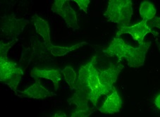
The core of this kit is a proprietary, cell-impermeant green fluorescent dye that selectively and stably incorporates into the outer leaflet of the plasma membrane lipid bilayer. The dye molecule is designed to be highly lipophilic, allowing it to partition rapidly into the membrane. Crucially, its chemical structure prevents it from flipping across the bilayer or being actively transported into the cell under normal conditions, ensuring the staining remains predominantly on the plasma membrane for extended periods (typically 30-60 minutes, depending on cell type and temperature). This results in a sharp, bright green fluorescent outline (with excitation/emission maxima typically around ~490/515 nm, compatible with standard FITC/GFP filter sets) that accurately defines cell contours and protrusions like microvilli or filopodia in real-time.
Key Features and Advantages of the TraKine™ Staining Kit
The TraKine™ Cell Plasma Membrane Staining Kit (KTC4001) is engineered for performance and convenience:
• High Specificity & Low Background: The dye exhibits strong preferential binding to the plasma membrane, yielding a crisp signal with minimal cytoplasmic or nuclear background.
• Live-Cell Compatibility: The staining protocol is fast and uses a dye concentration optimized for minimal phototoxicity and cellular perturbation, allowing for healthy, long-term live-cell imaging.
• Rapid and Simple Protocol: Staining is typically complete within 5-10 minutes at 37°C or 15-20 minutes at 4°C, involving a simple incubation step followed by a gentle wash. No complex preparation or waiting is required.
• Excellent Signal Stability: Once incorporated, the fluorescent signal remains stable for a sufficient duration to perform detailed microscopy or flow cytometry analysis without significant fading or redistribution.
• Versatility: The kit is suitable for staining a wide variety of adherent and suspension mammalian cell lines. It is ideal for confocal microscopy, widefield fluorescence microscopy, and flow cytometric analysis of cell surface labeling.
• Ready-to-Use Format: The kit includes a concentrated dye stock and a proven optimized staining buffer, simplifying workflow and ensuring consistent results across experiments.
Streamlined Staining Workflow for Immediate Results
The procedure is remarkably straightforward. Cells are grown under standard conditions. The concentrated dye is diluted in the provided assay buffer or an appropriate serum-free medium to create a working solution. This solution is added directly to the cells (covering the monolayer or suspending the cells) and incubated for a short period at the recommended temperature. After incubation, the staining solution is removed, and the cells are washed gently with buffer or pre-warmed culture medium to remove excess dye. The cells are then immediately ready for observation under a fluorescence microscope or analysis by flow cytometry. The entire process from dye addition to imaging can often be completed in under 20 minutes.
Diverse Applications in Cell Biology and Beyond
- Live-Cell Imaging and Morphology Studies: Clearly visualize cell shape, membrane ruffling, blebbing, and the dynamics of cellular protrusions in real-time, which is essential for research in cell migration, division, and apoptosis.
- Cell-Cell Interaction and Adhesion Assays: Precisely outline individual cells to study contact sites, synapse formation, or the boundaries in co-culture systems, providing critical spatial context.
- Membrane Integrity and Cytotoxicity Testing: Use the sharp membrane outline as a reference to assess membrane damage or pore formation induced by toxins, complement, or therapeutic agents. Disruption of the continuous fluorescent ring is a clear indicator of compromised membrane integrity.
- Flow Cytometry Gating and Analysis: Provide a bright, uniform surface stain that can be used in flow cytometry to accurately gate live cell populations based on size and membrane integrity, or to normalize other fluorescence signals to cell surface area.
- Reference Marker for Co-localization Studies: The clean plasma membrane stain serves as an excellent spatial reference point when co-staining with markers for intracellular organelles (e.g., mitochondria, lysosomes, nucleus) or specific surface proteins, enabling precise co-localization analysis.
- Educational Demonstrations: The simplicity and visual impact of the staining make it a powerful tool for teaching core concepts of cell structure and membrane biology in academic settings.
An Essential Tool for Visualizing Cellular Architecture
In the quest to understand cellular function, seeing is not just believing—it is understanding. The TraKine™ Cell Plasma Membrane Staining Kit (Green Fluorescence) empowers researchers with a reliable, efficient, and gentle method to illuminate the defining boundary of the cell. By delivering a bright, specific, and stable signal with minimal interference to cell physiology, this kit removes a common technical hurdle and opens a clear window into the dynamic world of the cell surface. It is an invaluable asset for any laboratory engaged in live-cell imaging, cytometric analysis, or fundamental cell biological research where the plasma membrane takes center stage.
Product Reference: KTC4001 – TraKine™ Cell Plasma Membrane Staining Kit (Green Fluorescence)
Learn more and order: https://www.abbkine.com/product/trakine-cell-plasma-membrane-staining-kit-green-fluorescence-ktc4001/


