


The Antibody That Unifies Your Epigenetics Workflow — Anti-Histone H3 Mouse Monoclonal Antibody (2D10)

The western-blot membrane sits on the light box, and the band at 15 kDa is so sharp it could cut glass. That band is Histone H3. For two decades it has been the quiet workhorse of every chromatin immunoprecipitation, every histone-modification western blot, every immunofluorescence panel that maps the geography of the nucleus. But the biochemist who first selected H3 as a loading control probably never imagined that the same antibody would one day be asked to perform in four different assays, across three mammalian species and a yeast model, while distinguishing genuine H3 from its variant cousins that can masquerade as the real thing. The gap between what H3 antibodies are asked to do and what most of them actually deliver is where Abbkine’s Anti‑Histone H3 Mouse Monoclonal Antibody (clone 2D10, catalog ABL1070) stakes its claim — and 12 peer‑reviewed publications suggest the claim holds up under scrutiny.
The antibody that makes this possible is not a polyclonal pool of uncertain composition. Clone 2D10 was generated by immunizing BALB/c mice with recombinant full‑length Histone H3 protein, then fusing the responding B cells with a myeloma partner to create a stable hybridoma that secretes a single immunoglobulin molecule targeting a single conserved epitope on the H3 polypeptide. That monoclonal architecture matters for a reason that datasheets rarely explain: polyclonal anti‑H3 antibodies recognize multiple epitopes scattered across the histone fold and the unstructured N‑terminal tail, and a fraction of those epitopes are shared with variant histones such as H3.3, H2A, or even H4. The resulting cross‑reactivity produces the blurry bands and foggy immunofluorescence images that reviewers notice. Abbkine’s quality‑control data specify a minimum signal‑to‑noise ratio of 22:1 in western blotting, and each production lot is tested for cross‑reactivity against non‑H3 histones. That level of validation is not a marketing ornament; it is the difference between a loading control you can defend in a revised manuscript and a loading control you quietly swap out after the third failed replicate.
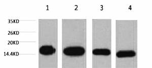
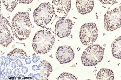
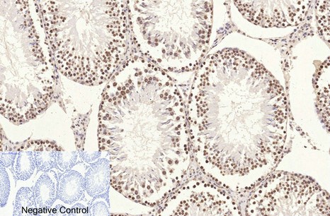
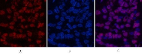
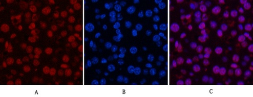
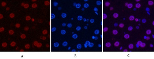
Abbkine recommends ABL1070 for western blot, immunohistochemistry on paraffin sections, immunofluorescence, and immunoprecipitation, with suggested starting dilutions of 1:2000–1:5000 for WB, 1:50–1:300 for IHC‑P, 1:100–1:500 for IF, and 1:200 for IP. These are realistic numbers. The product page shows a western blot image of HeLa cells, Raw264.7 macrophages, mouse brain, and rat brain, all processed at 1:5000, and the resulting bands are single, sharp, and positioned exactly at 15 kDa with no sign of saturation or ghosting. IHC images of paraffin‑embedded human uterus, mouse testis, and rat testis, each stained at 1:200, show nuclear‑restricted signal that follows the expected chromatin distribution without cytoplasmic haze. Immunofluorescence images of human liver‑cancer tissue, mouse liver, and rat liver, detected with a Cy3‑conjugated secondary antibody, resolve individual nuclei with clarity that permits co‑localization with DAPI without channel bleed‑through. An antibody that can pivot from denatured epitopes on a membrane to native epitopes in a tissue section without requiring separate lots for each application is an antibody whose epitope remains accessible across sample‑preparation chemistries — a property that polyclonal cocktails cannot guarantee.
Species reactivity extends to human, mouse, rat, and, critically, yeast. Most housekeeping‑protein antibodies ignore Saccharomyces cerevisiae entirely, which makes comparative‑biology workflows needlessly fragmented. ABL1070 detects yeast H3 with the same reagent that detects mammalian H3, enabling a chromatin‑biology laboratory to move from a yeast genetic screen to a mouse‑model validation to a human‑tissue confirmation without swapping loading controls mid‑project. That continuity removes a variable that is otherwise invisible but real: two different antibodies detecting the same target in two different species do not necessarily report the same abundance, because epitope‑presentation efficiency differs across proteomes. Removing that variable is not convenience; it is experimental hygiene.
The 12 publications that cite ABL1070 span a range that no single‑application antibody could accumulate. A 2017 study in Materials Science and Engineering: C (impact factor 27) used the antibody to normalize protein expression during an investigation of silica‑nanoparticle toxicity on adipogenic differentiation of human mesenchymal stem cells. A 2022 paper in Cell Communication and Signaling (impact factor 4) deployed ABL1070 for western‑blot normalization while dissecting the mechanism by which AKT inhibition triggers p53/SIRT6/PARP1‑dependent parthanatos. A colorectal‑cancer metastasis study in Experimental and Molecular Pathology (impact factor 2) relied on the antibody for loading‑control normalization during the characterization of formin‑like 3 expression. When a journal with an impact factor of 27 passes a loading‑control antibody through its peer‑review process without comment, the antibody has demonstrated a level of performance that no amount of in‑house validation can replicate.
Every researcher who works with histone‑H3 antibodies learns, sooner or later, that the loading‑control narrative conceals a deeper complexity. Histone H3 is not one protein; it is a family of replication‑dependent variants (H3.1, H3.2) and a replication‑independent variant (H3.3) that are deposited into chromatin by distinct chaperone pathways. Its N‑terminal tail carries a combinatorial code of post‑translational modifications — phosphorylation at Ser10, acetylation at Lys9 and Lys14, methylation at Lys4, Lys9, Lys27, Lys36, and Lys79 — that regulate transcription, DNA repair, and chromosome segregation. An antibody raised against a recombinant full‑length H3 protein, as 2D10 is, may or may not recognize the modified forms with equal affinity, and the answer to that question determines whether the loading‑control signal remains stable when a treatment alters the global modification state of chromatin. The product page does not claim that 2D10 is insensitive to modifications, which is the honest answer; most anti‑H3 antibodies are not. The appropriate response is the one that the best labs already practice: validate that total H3 abundance does not change under the specific experimental conditions before using it for normalization, and report that validation in the supplementary data. ABL1070 provides a reagent whose batch‑to‑batch consistency and defined epitope make such validation reproducible rather than reagent‑dependent.
Abbkine supplies the antibody as a liquid solution at 1 mg/mL in phosphate‑buffered saline, pH 7.4, with 0.02 % sodium azide as a preservative and 50 % glycerol as a cryoprotectant that depresses the freezing point and prevents immunoglobulin denaturation during storage at −20 °C. The vial ships on blue‑ice gel packs and remains stable for one year from the date of shipment. To recover product that may have adhered to the cap during transit, the protocol instructs users to centrifuge the vial after thawing and before opening — a small operational detail that preserves antibody mass. Aliquoting is advised to avoid repeated freeze‑thaw cycles, a standard precaution for any affinity‑purified monoclonal antibody. The product is for research use only and is not intended for diagnostic or therapeutic applications.
The internal‑control antibody that walks across all four core protein‑detection assays, recognizes yeast H3 with the same fidelity it brings to human, mouse, and rat samples, and arrives in a formulation stable at −20 °C for a year — that antibody changes the experimental design of an epigenetics project. It converts the internal control from a separate validation step into a continuous thread that runs through every assay and every species, reducing the uncontrolled variables that accumulate when different antibodies are substituted from experiment to experiment. The band at 15 kDa stays sharp. The nuclear signal stays nuclear. The yeast cross‑reactivity stays cross‑reactive. And the work of a chromatin‑biology laboratory becomes a little easier to reproduce.
Explore specifications, view representative images, and place your order here: https://www.abbkine.com/product/anti-histone-h3-mouse-monoclonal-antibody-2d10-abl1070/


