| Product name | Anti-Histone H3 Mouse Monoclonal Antibody (2D10) |
| Immunogen | Recombinant Protein |
| Host | Mouse |
| Reactivity | Human, Mouse, Rat, Yeast |
| Applications | IF, IHC, IP, WB |
| Applications notes | Optimal working dilutions should be determined experimentally by the investigator. Suggested starting dilutions are as follows: WB (1:2000-1:5000), IHC-P (1:50-1:300), IF (1:100-1:500), IP (1:200). |
| Clonality | Monoclonal |
| Preparation method | The antibody was affinity-purified from mouse ascites by affinity-chromatography using specific immunogen |
| Alternative | HIST1H3A; H3FA; HIST1H3B; H3FL; HIST1H3C; H3FC; HIST1H3D; H3FB; HIST1H3E; H3FD; HIST1H3F; H3FI; HIST1H3G; H3FH; HIST1H3H; H3FK; HIST1H3I; H3FF; HIST1H3J; H3FJ; Histone H3.1; Histone H3/a; Histone H3/b; Histone H3/c; Histone H3/d; Histone H3/f; Histone H3 |
| Formulation | Liquid solution |
| Storage buffer | Liquid in PBS, pH 7.4, containing 0.02% Sodium Azide as preservative and 50% Glycerol. |
| Storage instructions | Stable for one year at -20°C from date of shipment. For maximum recovery of product, centrifuge the original vial after thawing and prior to removing the cap. Aliquot to avoid repeated freezing and thawing. |
| Shipping | Gel pack with blue ice. |
| Precautions | The product listed herein is for research use only and is not intended for use in human or clinical diagnosis. Suggested applications of our products are not recommendations to use our products in violation of any patent or as a license. We cannot be responsible for patent infringements or other violations that may occur with the use of this product. |
| Background | Histone H3 is one of the five main histone proteins involved in the structure of chromatin in eukaryotic cells. Core component of nucleosome. Nucleosomes wrap and compact DNA into chromatin, limiting DNA accessibility to the cellular machineries which require DNA as a template. Histones thereby play a central role in transcription regulation, DNA repair, DNA replication and chromosomal stability. |
| Gene ID | 8350/8351/8352/8353/8354/8355/8356/8357/8358/8968 |
| Alternative | HIST1H3A; H3FA; HIST1H3B; H3FL; HIST1H3C; H3FC; HIST1H3D; H3FB; HIST1H3E; H3FD; HIST1H3F; H3FI; HIST1H3G; H3FH; HIST1H3H; H3FK; HIST1H3I; H3FF; HIST1H3J; H3FJ; Histone H3.1; Histone H3/a; Histone H3/b; Histone H3/c; Histone H3/d; Histone H3/f; Histone H3 |
| Accession | P68431/Q71DI3/P84243 |
| Observed Band(KD) | 15 |
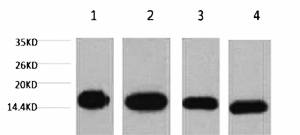
Fig.1. Western blot analysis of 1) Hela, 2) Raw, 3) mouse brain tissue, 4) rat brain tissue, diluted at 1:5000.
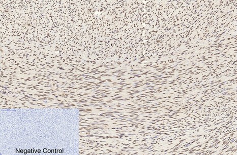
Fig.2. Immunohistochemical analysis of paraffin-embedded human uterus tissue. 1, Histone H3 Monoclonal Antibody (2D10) was diluted at 1:200 (4°C, overnight). 2, Sodium citrate pH 6.0 was used for antibody retrieval (>98°C, 20min). 3, secondary antibody was diluted at 1:200 (room temperature, 30min). Negative control was used by secondary antibody only.
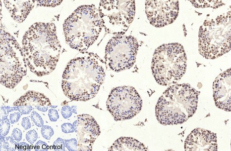
Fig.3. Immunohistochemical analysis of paraffin-embedded mouse testis tissue. 1, Histone H3 Monoclonal Antibody (2D10) was diluted at 1:200 (4°C, overnight). 2, Sodium citrate pH 6.0 was used for antibody retrieval (>98°C, 20min). 3, secondary antibody was diluted at 1:200 (room temperature, 30min). Negative control was used by secondary antibody only.
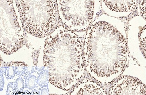
Fig.4. Immunohistochemical analysis of paraffin-embedded rat testis tissue. 1, Histone H3 Monoclonal Antibody (2D10) was diluted at 1:200 (4°C, overnight). 2, Sodium citrate pH 6.0 was used for antibody retrieval (>98°C, 20min). 3, secondary antibody was diluted at 1:200 (room temperature, 30min). Negative control was used by secondary antibody only.

Fig.5. Immunofluorescence analysis of human liver cancer tissue. 1, Histone H3 Monoclonal Antibody (2D10) (red) was diluted at 1:200 (4°C, overnight). 2, Cy3 Labeled secondary antibody was diluted at 1:300 (room temperature, 50min). 3, Picture B: DAPI (blue) 10min. Picture A: Target. Picture B: DAPI. Picture C: merge of A+B.
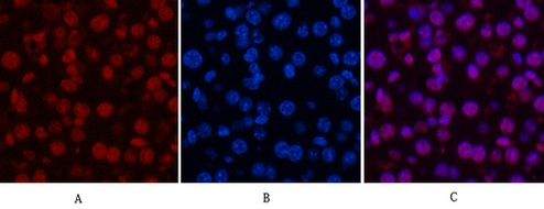
Fig.6. Immunofluorescence analysis of mouse liver tissue. 1, Histone H3 Monoclonal Antibody (2D10) (red) was diluted at 1:200 (4°C, overnight). 2, Cy3 Labeled secondary antibody was diluted at 1:300 (room temperature, 50min). 3, Picture B: DAPI (blue) 10min. Picture A: Target. Picture B: DAPI. Picture C: merge of A+B.
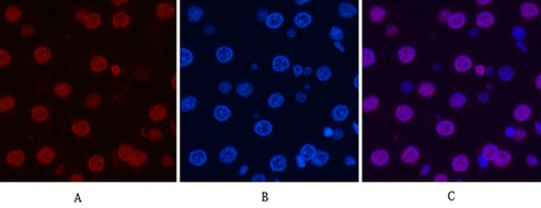
Fig.7. Immunofluorescence analysis of rat liver tissue. 1, Histone H3 Monoclonal Antibody (2D10) (red) was diluted at 1:200 (4°C, overnight). 2, Cy3 Labeled secondary antibody was diluted at 1:300 (room temperature, 50min). 3, Picture B: DAPI (blue) 10min. Picture A: Target. Picture B: DAPI. Picture C: merge of A+B.
Author:Yang X, Liu X J, Li Y Y, et al Publication name:Materials Science and Engineering: C IF:27
Author:Mi, Ai, et al Publication name:Food & Function IF:6
Author:Niu, Xuan, et al Publication name:Arteriosclerosis, Thrombosis, and Vascular Biology (2017): ATVBAHA-116 IF:5
Author:Liu, Jialin, et al. Publication name: Biomedical and Environmental Sciences IF:4.1
Author:Zhang Y, Zhang C, Li J, Jiang M, Guo S, Yang G, Zhang L, Wang F, Yi S, Wang J, Fu Y, Zhang Y Publication name:Cell Commun Signal IF:4
Author:Zhang Z, Bu X, Chen H, et al Publication name:International Journal of Molecular Medicine IF:3
Author:Ma F, Wang L and Wang Y Planta, 2018 Publication name: IF:3
Author:Luo Q, Li W, Li M, et al Publication name:Life sciences IF:3
Author:Li G, Jia Z, Cao Y, et al Publication name:Neurochemical research IF:3
Author:P Hu, C Zhu Publication name:ENDOCR METAB IMMUNE IF:2
Author:Zeng Y F, Xiao Y S, Lu M Z, et al Publication name:Experimental and molecular pathology IF:2
Author:Chen, Liu, et al. Publication name: bioRxiv IF:
You must be logged in to post a review.


Reviews
There are no reviews yet.