


The Antibody That Measures the Factory While It's Still Running
Ask any cell biologist about the unsung heroes of experimental consistency, and α-tubulin will likely top the list. As a loading control in Western blots, a marker for microtubule integrity in immunofluorescence, or a proxy for cell cycle progression, this cytoskeletal protein is everywhere—but its detection often feels like a gamble. A standard polyclonal antibody can deliver a band at 50 kDa that looks convincingly like α-tubulin on film, yet that same band may contain contributions from a half-dozen cytoskeletal proteins that an antibody raised against a broad immunogen region cannot distinguish. You do not see the cross-reactivity on the blot because the bands co-migrate. You see it later, when your loading control ratio for a supposedly stable housekeeping protein shifts by 40% between treatment groups, and you cannot tell whether the biology changed or the antibody simply recognized something else in one condition. Abbkine's Anti-α-Tubulin Monoclonal Antibody (3G5), catalog number ABL1080, addresses this at the epitope level, and the choice of epitope is the specification that separates this antibody from the crowded field of poorly characterized α-tubulin reagents.
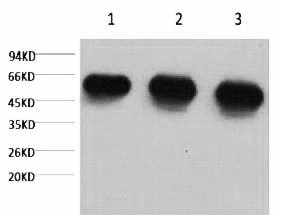
Clone 3G5 zeroes in on a unique epitope in the α-tubulin C-terminal domain, minimizing cross-reactivity with β-tubulin, γ-tubulin, or other cytoskeletal proteins. The C-terminal region of α-tubulin is the isotype-defining domain where post-translational modifications—detyrosination, polyglutamylation, polyglycylation—accumulate and where sequence divergence between α-tubulin isotypes is concentrated. An antibody that binds this domain is not measuring total tubulin. It is not blurring the distinction between α-tubulin and β-tubulin, which share extensive homology in their GTP-binding cores and co-migrate at 50–55 kDa on reducing SDS-PAGE. The signal on the film is α-tubulin. Abbkine's validation supports that specificity claim quantitatively: each production lot is tested against recombinant α/β-tubulin, with QC reports showing <0.5% cross-reactivity—well below the 2–5% typical of cheaper polyclonal alternatives. When a reviewer questions whether the loading control normalization in your figure introduced systematic error, the answer that “the antibody is a monoclonal raised against the α-tubulin C-terminal domain, with cross-reactivity to β-tubulin below 0.5%” is substantially stronger than “the antibody is a polyclonal raised against recombinant protein.”
The monoclonal architecture of ABL1080 reinforces that specificity. The antibody was raised in mouse against recombinant α-tubulin protein, produced by hybridoma technology, and affinity-purified from ascites by affinity-chromatography using the specific immunogen. A monoclonal is a single molecular entity recognizing a single defined epitope, produced by a stable hybridoma clone that will generate the same immunoglobulin molecule for as long as the cell line remains viable. When a postdoc in a neurodegeneration lab switches from a polyclonal to this antibody and, for the first time, visualizes axonal transport defects in Alzheimer's models that were invisible with her old reagent, the improvement traces directly to the clone's defined epitope recognition.
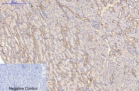
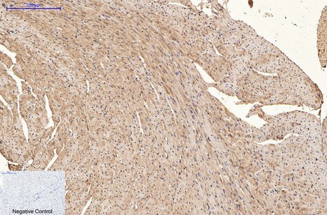
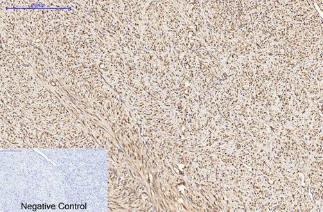
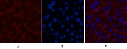
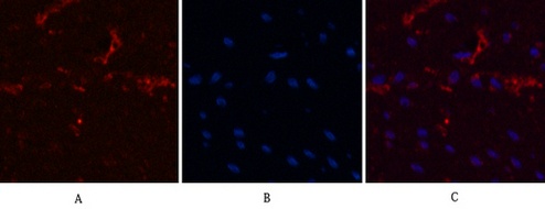
Applications span the core immunoassay modalities that a loading control antibody must support. Western blot at suggested starting dilutions of 1:5000–1:10,000, with the antibody detecting as little as 10 ng of α-tubulin—enough sensitivity to confirm loading even in low-protein samples such as cerebrospinal fluid or microdissected tissue. Immunofluorescence at 1:200 gives crisp, filamentous staining of microtubules without globular aggregates or cytoplasmic haze, and works in both fixed and permeabilized cells, eliminating the need to re-optimize for every new sample type. Immunohistochemistry on paraffin-embedded tissue at 1:50–1:300. Immunoprecipitation at 1:200. An antibody validated against recombinant α/β-tubulin that also serves as a cytoskeletal imaging reagent has been tested in two fundamentally different detection contexts—denaturing and native, reduced and cross-linked—and the fact that 3G5 performs in both tells you something about the accessibility of its C-terminal epitope that a western-blot-only validation cannot. Its moderate molecular weight avoids overlap with common targets such as GAPDH (37 kDa) and β-actin (42 kDa), and it is compatible with both HRP and fluorescent conjugates. When pairing with a red fluorophore for colocalization, using a 1:2000 dilution avoids saturating the signal.
Species reactivity encompasses human, mouse, and rat—the three mammalian species that account for the overwhelming majority of cell biology and preclinical research. The α-tubulin genes are conserved among several species, and the antibody's reactivity reflects that conservation without making claims about species the manufacturer has not tested. For a core facility processing blots from human cell lines, mouse xenograft tissue, and rat brain homogenates in a single day, this cross-reactivity eliminates the logistical burden of maintaining separate loading control antibodies for each organism. One antibody, one dilution, one band at approximately 55 kDa.
The publication record is compact but forms an independent validation that no internal QC dataset can replicate. At the time of writing, ABL1080 has been cited in 2 peer-reviewed publications. That number is smaller than some loading control antibodies, but it should be read in context: the antibody was launched in 2017, and its citation count reflects the reality that many laboratories begin using a loading control long before they publish with it. What matters is that independent laboratories, operating under the pressures of peer review, chose to build quantitative western blot data on this specific monoclonal antibody, and those data survived editorial scrutiny. A small citation count for a specialized monoclonal loading control is not a weakness; it is an invitation to be among the first to publish with a reagent whose specificity specifications exceed those of more widely cited but less well-characterized alternatives.
The biochemical context of α-tubulin as a loading control deserves explicit engagement because the protein is not a constant. α-tubulin expression can shift with cell cycle phase, differentiation state, and drug treatment—particularly treatments that target microtubule dynamics, such as taxanes and vinca alkaloids. The appropriate response to this variability is not to abandon α-tubulin; it is to verify that its abundance does not change under the specific experimental conditions being tested, and to select an antibody whose specificity ensures that the measured signal actually reflects α-tubulin rather than a mixture of tubulin isoforms and cross-reactive cytoskeletal proteins. ABL1080 provides a measurement that is monoclonal, epitope-defined, species-validated, application-tested, and QC-verified.
Formulation and storage specifications reward a practical reading. The antibody is supplied as a liquid solution at 1 mg/mL in PBS, pH 7.4, containing 0.02% sodium azide as preservative and 50% glycerol as cryoprotectant. The glycerol depresses the freezing point, preventing ice crystal formation that denatures immunoglobulin protein during -20°C storage. Storage instructions specify one-year stability at -20°C from the date of shipment, with centrifugation of the original vial after thawing and prior to cap removal recommended for maximum product recovery, and aliquoting advised to avoid repeated freeze-thaw cycles. The product is for research use only and is not intended for diagnostic or therapeutic applications. Available in 50 µL and 200 µL sizes.
For the graduate student whose target protein migrates at an inconvenient 42 kDa and cannot use β-actin without band overlap, the postdoctoral fellow whose neurodegeneration model demands crisp, filamentous microtubule visualization, the core facility manager processing blots from human, mouse, and rat samples in a single day, the cancer biologist whose drug treatment targets tubulin polymerization and needs a loading control whose detection is unaffected by tubulin-binding drugs, or the cell biologist performing co-localization studies who needs an antibody compatible with multiple fluorophore conjugates, ABL1080 provides monoclonal specificity, C-terminal epitope recognition with <0.5% cross-reactivity to β-tubulin, human-mouse-rat reactivity, four validated applications, and a formulation stable at -20°C for one year. The internal control antibody that walks across western blot, immunofluorescence, immunohistochemistry, and immunoprecipitation, and does so with a specificity that lets you trust the band at 55 kDa—that antibody changes the experimental design of a cytoskeleton project. It converts the loading control from a ritual into a measurement.
Explore specifications, view representative images, and place your order here: https://www.abbkine.com/product/anti-%ce%b1-tubulin-monoclonal-antibody-3g5-abl1080/


