


The $69 ECL Substrate That Tracks A 0.5-Picogram Band For A Full Hour
A postdoctoral fellow in a mitochondrial disease lab once told me that the moment she truly grasped the difference between a quality chemiluminescent substrate and a generic one was not when she imaged a high‑abundance loading control. It was when she had to detect the translocase of the inner mitochondrial membrane 23 (TIM23) from a primary neuron lysate yielding only 15 µg of total protein. The first blot, developed with the inexpensive substrate her purchasing department had found, showed a blank lane where the 22 kDa band should have been. She doubled the protein load, optimized the transfer, and repeated the blot. A faint band appeared. The reviewer requested quantification, and the signal‑to‑noise ratio fell below three—statistically indistinguishable from background. She switched substrates. The same primary antibody, the same secondary antibody, the same membrane, and the TIM23 band appeared with an intensity that allowed densitometry. The difference was not the antibody. It was the duration and intensity of the light that reached the CCD chip, and the substrate that generated it had failed before the camera finished initializing.
Western blotting remains an indispensable technique for protein detection, but the quality of the ECL substrate can determine the success or failure of an experiment. For researchers pursuing ultra‑sensitive, reliable detection of low‑abundance proteins, Abbkine‘s SuperKine™ West Femto Maximum Sensitivity Substrate (BMU102‑EN) is engineered to push the limits, generating intense, stable chemiluminescent signals that enable accurate quantification of proteins that conventional substrates might miss. This substrate has now accumulated over 8,900 product page views and 20 peer‑reviewed citations. One of those citations appears in Journal of Hospital Infection (IF 6.9), where the kit was deployed within a universal Western blot toolkit for comparative efficacy evaluation of disinfectants against SARS‑CoV‑2. Another study on the molecular mechanisms of cisplatin‑induced apoptosis used BMU102‑CN for HRP‑based detection of Bax, Bcl‑2, and phosphorylated NF‑κB p65, requiring linear signal across target abundances that differed by more than an order of magnitude. A third publication in PLOS ONE examined astragaloside IV attenuation of high‑glucose‑induced peritoneal fibrosis, relying on the substrate to detect low‑abundance ENKUR and PI3K/Akt pathway components in human peritoneal mesothelial cells. These are not citations in journals that accept anything with a p‑value. They represent independent laboratories that prepared their own blots, used their own antibodies, operated their own imaging equipment, and submitted their own figures for peer review with this specific substrate listed in the methods section.
The chemistry that enables this sensitivity is built on innovation and optimization of the classic peroxidase substrate system, engineered to generate intense light output that does not fade before the detector can capture it. The working solution is prepared by mixing equal volumes of the two provided components, and 20 citations from independent laboratories confirm that the luminescence persists long enough for multiple exposures, CCD camera focusing adjustments, and optimal image capture. Pour the substrate onto the membrane, incubate, drain the excess, and expose. The signal that reaches the film or digital imager is not a transient flash that decays before the instrument finishes its initialization routine; it is a stable, prolonged emission that allows the researcher to capture the image at the exposure time that places all bands of interest within the linear dynamic range of the detection system.
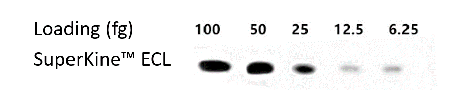
The sensitivity specification is calibrated to the biological reality of low‑abundance protein detection. The product page describes Femto‑level maximum sensitivity as a game‑changer for detecting trace amounts of target proteins. The substrate detects as little as 0.5 pg of protein, a sensitivity that places it in the same category as premium femto‑grade substrates costing three to four times as much, and provides a high‑sensitivity color developing solution to enhance the chemical signal of immunological experiments.
Signal duration is the counter‑specification that matters most for a femto‑grade substrate. Multiple data sheets confirm that the signal remains stable for extended periods, allowing for multiple exposures, CCD camera focusing adjustments, and optimal image capture. The substrate‘s intense signal output and duration enables detection of picogram amounts of antigen with photographic or other imaging methods, though it is best to perform compression or imaging within 30 minutes of color development. This is not a substrate that demands the operator race from the darkroom to the imager before the light dies. It is a substrate that gives the operator time to optimize the capture, verify that bands fall within the linear range, and re‑image if the first exposure is not optimal.
Compatibility is a specification that matters most to the researcher who processes 30 Western blots per month and cannot afford to re‑optimize protocols every time a new substrate arrives. The SuperKine™ West Femto Substrate integrates seamlessly into standard Western blot workflows, requiring no special equipment and no complex protocol adjustments. It is compatible with all common membrane types including PVDF and nitrocellulose, and with HRP‑conjugated secondary antibodies. The working solution is prepared at a 1:1 ratio, the same protocol that governs virtually every other ECL substrate on the market, which means laboratories already using ECL detection can switch to BMU102‑EN without re‑optimizing blocking conditions, washing steps, or exposure times.
Economic accessibility distinguishes BMU102‑EN from the premium femto‑grade substrates whose sensitivity it matches. The product is priced at ‑72. The product page emphasizes that Abbkine‘s commitment to making high‑quality research tools accessible means researchers do not have to sacrifice sensitivity or reliability to stay within budget. For laboratories in countries where research budgets are constrained, the difference between 250 for a femto‑grade substrate is the difference between running the confirmatory replicates a reviewer requested and omitting them because the substrate budget was exhausted.
The stability and storage specifications are practical and undemanding: stable for one year at 4–8°C from date of shipment, exposure to intense light such as direct sun can harm the working solution, but short‑term exposure to typical laboratory lighting will not. Shipping occurs on gel packs with blue ice, maintaining the cold chain from the manufacturer‘s warehouse to the laboratory freezer. The 100 mL size consists of 50 mL of Reagent A and 50 mL of Reagent B, yielding 100 mL of working solution. The protocol—mix the two substrate components at a 1:1 ratio, incubate the blot for 1–5 minutes, drain excess reagent, cover with plastic wrap, and expose—is the same protocol that governs every other ECL substrate on the market, which means switching to BMU102‑EN does not require re‑writing the laboratory‘s Western blot standard operating procedure.
The broader technological context makes the case for selecting a femto‑grade ECL substrate with long signal duration increasingly urgent as quantitative Western blotting standards tighten. A growing number of journals and funding agencies require quantitative Western blot data with demonstrable linear dynamic range, and the signal decay kinetics of the detection substrate become an analytical variable rather than a technical footnote. A substrate that loses 80% of its signal within five minutes forces the user to capture the image at a single time point that may fall outside the linear range of the detection system for either the highest‑ or lowest‑abundance bands on the membrane. A substrate whose signal remains stable for extended periods allows the user to capture multiple exposures, verify that band intensities fall within the linear range, and select the exposure that provides accurate quantification across all bands of interest.
For the postdoctoral fellow whose low‑abundance mitochondrial protein has resisted detection through four different primary antibodies and three different transfer buffers, the graduate student whose phosphorylated signaling protein represents less than 0.001% of total cellular protein and whose entire experiment hinges on visualizing that single band, the core facility manager who must standardize on a single substrate across diverse user projects without compromising detection sensitivity, the principal investigator submitting grant renewal data that must survive reviewer scrutiny of every band on every blot, and the researcher in any laboratory anywhere who has watched a chemiluminescent signal fade before the CCD camera finished initializing, BMU102‑EN provides femtogram‑level sensitivity, a signal duration that supports multiple exposures, drop‑in compatibility with existing ECL protocols, 20 peer‑reviewed publication citations, one‑year stability at 4–8°C, and a $69 price for 100 mL. The signal you have been losing to substrate decay before the detector even warms up can now be captured, quantified, and published.
Explore specifications, view representative data, and place your order here: https://www.abbkine.com/product/superkine-west-femto-maximum-sensitivity-substrate-bmu102-en/


