




p38 MAPK: The Cellular Stress Sentinel – How the Abbkine p38 Polyclonal Antibody (ABP0150) Unlocks Insights into Inflammation, Cancer, and Beyond
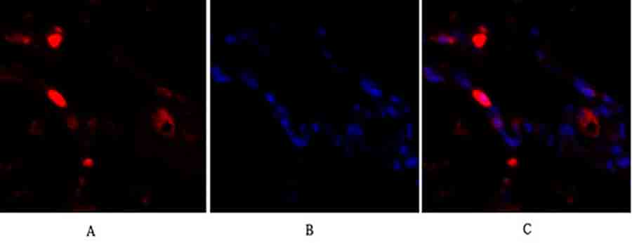
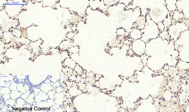
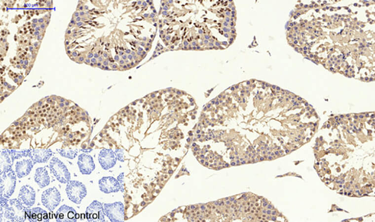
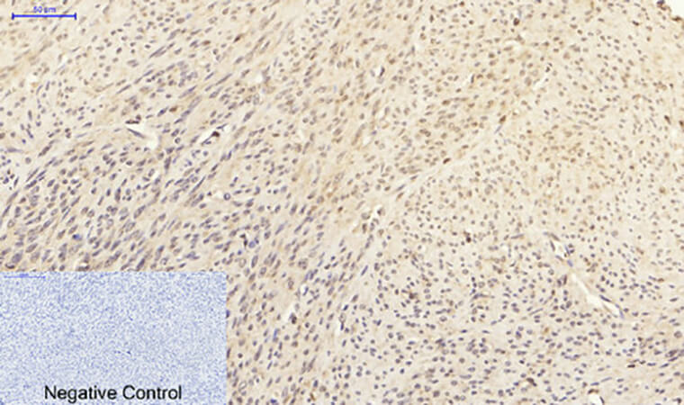
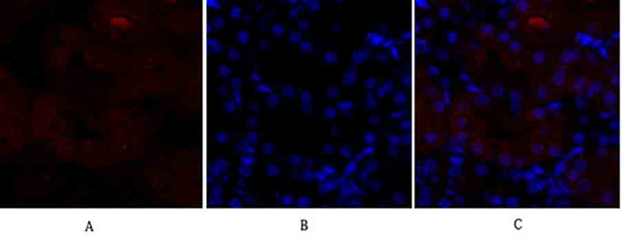
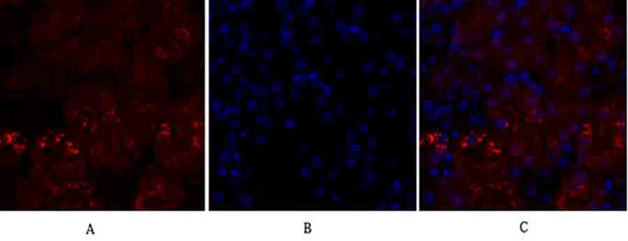
When a cell faces the onslaught of inflammatory cytokines, osmotic shock, UV radiation, or genotoxic stress, a molecular alarm system is triggered—a cascade of phosphorylation events that converge on a central kinase known as p38 mitogen-activated protein kinase (MAPK). This evolutionarily conserved stress sensor, first identified as a target of pyridinyl imidazole inhibitors like SB203580, is far more than a simple signaling node; it is a master regulator of cellular fate, dictating responses ranging from apoptosis and cell cycle arrest to cytokine production and differentiation . Dysregulation of p38 signaling underpins a vast spectrum of human pathologies, from rheumatoid arthritis and inflammatory bowel disease to neurodegenerative disorders and metastatic cancer . Consequently, precisely detecting and quantifying p38 protein expression and activation status is a cornerstone of modern molecular pathology, drug discovery, and basic research into stress adaptation. However, p38 presents unique detection challenges: it exists as four isoforms (p38α, β, γ, δ) with overlapping yet distinct functions, undergoes rapid phosphorylation-dependent activation, and shuttles between the cytoplasm and nucleus upon stimulation . The Abbkine p38 Polyclonal Antibody (ABP0150) is engineered to meet these challenges, providing researchers with a high-affinity, rabbit-derived polyclonal reagent rigorously validated for Western blot (WB), immunohistochemistry (IHC-P), immunofluorescence (IF), immunoprecipitation (IP), and ELISA, with confirmed reactivity against human, mouse, and rat p38 MAPK . Whether you are mapping the inflammatory landscape of a tumor microenvironment, dissecting neuronal death pathways in Alzheimer's models, or screening novel p38 inhibitors for autoimmune therapy, this antibody delivers the specificity, sensitivity, and versatility required to generate robust, reproducible data across diverse experimental systems.
The p38 Signaling Nexus: From Stress Response to Therapeutic Target
p38 MAPK (UniProt Q16539, gene symbol MAPK14) is a ~41 kDa serine/threonine kinase belonging to the MAPK superfamily, which also includes ERK1/2 and JNK . Four isoforms—p38α (MAPK14), p38β (MAPK11), p38γ (MAPK12/ERK6), and p38δ (MAPK13/SAPK4)—are encoded by distinct genes and exhibit tissue-specific expression and functional specialization . p38α is the most ubiquitously expressed and best-characterized isoform, serving as the primary mediator of cellular stress responses . Activation occurs through dual phosphorylation of the conserved Thr-Gly-Tyr motif (Thr180/Tyr182 in p38α) by upstream kinases MKK3 and MKK6, which are themselves activated by a cascade of MAP3Ks in response to diverse stimuli . Once activated, p38 phosphorylates an astonishing array of over 200 substrates, including transcription factors (ATF2, MEF2C, p53, CHOP), kinases (MAPKAPK2/3, MSK1/2), and proteins regulating mRNA stability (TTP, HuR) and translation . This broad substrate specificity allows p38 to orchestrate inflammatory cytokine production (TNF-α, IL-1β, IL-6), cell cycle arrest at G1/S and G2/M checkpoints, apoptosis in response to severe stress, and differentiation of immune cells, neurons, and adipocytes . In cancer, p38 plays a context-dependent dual role: it can act as a tumor suppressor by inducing apoptosis and senescence in premalignant cells, yet in established tumors, it often promotes survival, invasion, and therapy resistance by driving pro-inflammatory and pro-angiogenic signals . In the nervous system, sustained p38 activation contributes to synaptic dysfunction, neuroinflammation, and neuronal death in Alzheimer's, Parkinson's, and ALS . Thus, reliable detection of total p38 protein—and its phosphorylated active form—is indispensable for understanding disease mechanisms, validating drug targets, and stratifying patients for targeted therapies.
Antibody Specifications: A Versatile Tool for Multi-Dimensional p38 Analysis
The Abbkine p38 Polyclonal Antibody (ABP0150) is an affinity-purified rabbit polyclonal antibody generated using a synthetic peptide derived from a conserved region of human p38 MAPK, ensuring broad recognition of p38 isoforms across multiple species . Key characteristics include:
• Host Species: Rabbit – ideal for multiplex staining with mouse-derived primary antibodies in IHC/IF and compatible with most secondary detection systems.
• Reactivity: Confirmed for human, mouse, and rat p38 MAPK, enabling translational studies from rodent models to human clinical samples .
• Applications: Validated for Western blot (WB), immunohistochemistry on paraffin-embedded sections (IHC-P), immunofluorescence (IF), immunoprecipitation (IP), and ELISA . Recommended starting dilutions are:
◦ Western blot: 1:500 to 1:2000

◦ Immunohistochemistry (IHC-P): 1:100 to 1:300

◦ Immunofluorescence (IF): 1:50 to 1:200

◦ ELISA: 1:10000

◦ Immunoprecipitation (IP): 1:10 to 1:200
• Clonality: Polyclonal – recognizes multiple epitopes on the p38 protein, enhancing detection sensitivity and robustness, particularly for denatured samples in WB and fixed tissues in IHC .
• Target Specificity: Detects total p38 protein (primarily p38α, with likely cross-reactivity to β and γ isoforms based on sequence homology); does not distinguish phosphorylated (active) from non-phosphorylated forms—for phospho-specific detection, a companion phospho-p38 antibody is required .
• Formulation: Supplied as a liquid solution at 1 mg/mL in PBS (pH 7.4) containing 0.5% BSA (stabilizer), 0.02% sodium azide (preservative), and 50% glycerol for long-term storage .
• Storage: Stable for one year at –20°C; avoid repeated freeze-thaw cycles by aliquoting upon receipt .
Five Critical Research Applications Enabled by the Abbkine p38 Antibody
Research Domain Specific Investigation How ABP0150 Provides the Answer
Inflammatory Disease & Autoimmunity Mapping p38 activation in synovial tissue from rheumatoid arthritis patients or intestinal biopsies from Crohn's disease to correlate kinase activity with disease severity and response to biologics (e.g., anti-TNF therapy). Perform IHC-P on FFPE synovium/colon sections using ABP0150 (1:200) to quantify total p38 expression; combine with phospho-p38 staining to assess activation status .
Cancer Biology & Tumor Microenvironment Assessing p38 expression and subcellular localization in triple-negative breast cancer (TNBC) or glioblastoma to determine its role in therapy resistance, epithelial-mesenchymal transition (EMT), and inflammatory niche formation. Use IF on cancer cell lines or tissue microarrays to visualize cytoplasmic vs. nuclear p38; co-stain with phospho-p38 and proliferation markers (Ki-67) to link signaling to tumor growth .
Neurodegeneration & Brain Injury Investigating p38-mediated neuroinflammation in Alzheimer's mouse models (e.g., APP/PS1) or after traumatic brain injury, focusing on microglial activation and tau hyperphosphorylation. Conduct Western blot on hippocampal lysates (1:1000) to measure total p38 levels; combine with IP using ABP0150 to pull down p38 complexes for phospho-tau analysis .
Cardiovascular Stress & Ischemia Evaluating p38 activation in cardiomyocytes after myocardial infarction or in endothelial cells under shear stress, linking oxidative stress to apoptosis and cardiac remodeling. Apply IHC-P on heart tissue sections (1:150) to detect p38 in infarct border zones; use ELISA (1:10000) on serum/plasma to quantify soluble p38 as a potential biomarker of cardiac stress .
Drug Discovery & Pharmacology Screening novel p38 inhibitors in LPS-stimulated macrophages by monitoring downstream phosphorylation of MAPKAPK2 or cytokine production (TNF-α, IL-6). Perform Western blot on cell lysates treated with inhibitor candidates; use ABP0150 to confirm total p38 loading control while probing with phospho-p38 and phospho-MAPKAPK2 antibodies .
Step-by-Step Protocols for Optimal p38 Detection
① Western Blot for Total p38 Protein
• Sample preparation: Lyse cells or tissues in RIPA buffer supplemented with protease and phosphatase inhibitors (sodium orthovanadate, β-glycerophosphate, PMSF). For phospho-protein analysis, include PhosSTOP tablets.
• Gel electrophoresis: Load 20–40 µg total protein on a 10–12% SDS-PAGE gel. Include a positive control (e.g., lysate from HeLa cells treated with anisomycin or UV to activate p38).
• Transfer: Transfer to PVDF membrane at 100 V for 60–90 min in ice-cold transfer buffer.
• Blocking: Block with 5% BSA or non-fat milk in TBST for 1 h at room temperature (BSA is preferred for subsequent phospho-blotting).
• Primary antibody: Incubate with Abbkine p38 antibody (ABP0150) diluted 1:1000 in blocking buffer overnight at 4°C with gentle agitation.
• Washing: Wash 3 × 10 min with TBST.
• Secondary antibody: Incubate with HRP-conjugated anti-rabbit IgG (1:5000) for 1 h at room temperature.
• Detection: Develop with enhanced chemiluminescence (ECL) substrate and image. Expected band at ~41 kDa; minor bands may represent isoforms or degradation products.
② Immunohistochemistry (IHC-P) on Formalin-Fixed Paraffin-Embedded Tissues
• Deparaffinization & antigen retrieval: Bake slides at 60°C for 1 h, deparaffinize in xylene, rehydrate through graded ethanol. Perform heat-induced epitope retrieval (HIER) in citrate buffer (pH 6.0) or EDTA buffer (pH 9.0) using a pressure cooker or microwave for 20 min.
• Peroxidase quenching: Block endogenous peroxidase with 3% H₂O₂ in methanol for 10 min.
• Blocking: Block non-specific binding with 5% normal goat serum for 30 min.
• Primary antibody: Apply ABP0150 at 1:200 dilution in antibody diluent and incubate overnight at 4°C in a humidified chamber.
• Detection: Use a polymer-based HRP detection system (e.g., DAB) followed by hematoxylin counterstaining.
• Interpretation: Cytoplasmic and/or nuclear staining is expected depending on cellular activation state; endothelial cells, macrophages, and epithelial cells often show strong p38 expression.
③ Immunofluorescence (IF) for Subcellular Localization
• Cell culture & stimulation: Seed cells on coverslips, treat with stress inducers (e.g., 10 ng/mL anisomycin for 30 min, 1 M sorbitol for 1 h, or 10 ng/mL TNF-α for 15 min) to trigger p38 nuclear translocation.
• Fixation & permeabilization: Fix with 4% paraformaldehyde (PFA) for 15 min, permeabilize with 0.1% Triton X-100 for 10 min.
• Blocking: Block with 5% BSA + 0.1% Tween-20 for 1 h.
• Primary antibody: Incubate with ABP0150 (1:100) and a mouse anti-phospho-p38 antibody overnight at 4°C to co-stain total and active p38.
• Secondary antibodies: Use Alexa Fluor 488-conjugated anti-rabbit and Alexa Fluor 594-conjugated anti-mouse (1:500) for 1 h at room temperature in the dark.
• Mounting & imaging: Mount with DAPI-containing medium and image using a confocal microscope; nuclear accumulation of p38 upon stress is a key readout.
④ Immunoprecipitation (IP) for Protein Complex Analysis
• Lysate preparation: Lyse cells in non-denaturing IP buffer (e.g., 50 mM Tris-HCl pH 7.4, 150 mM NaCl, 1% NP-40) with protease/phosphatase inhibitors.
• Pre-clearing: Incubate lysate with protein A/G beads for 1 h at 4°C to reduce non-specific binding.
• Immunoprecipitation: Add 2–5 µg of ABP0150 to 500 µg of lysate and incubate overnight at 4°C with rotation. Then add protein A/G beads for 2 h.
• Washing & elution: Wash beads 3–4 times with IP buffer, elute proteins with 2× Laemmli buffer by boiling for 5 min.
• Analysis: Run eluate on SDS-PAGE and probe for p38-interacting proteins (e.g., MK2, TAB1, MKK3) or post-translational modifications.
Troubleshooting Guide for Reliable p38 Staining
Issue Potential Cause Recommended Solution
Weak or no signal in Western blot Insufficient protein loading (p38 is abundant but may be poorly extracted); over-transfer; inefficient antigen retrieval for IHC. Load 30–50 µg of total protein; optimize transfer time (90 min for 41 kDa); for IHC, try high-pH EDTA retrieval (pH 9.0).
High background in IHC/IF Non-specific binding of primary/secondary antibody; endogenous peroxidase/alkaline phosphatase activity; incomplete blocking. Use species-appropriate normal serum blocking; extend H₂O₂ quenching to 15 min; titrate primary antibody (try 1:400–1:800).
Multiple bands in Western blot Cross-reactivity with other MAPK family members (ERK, JNK) or p38 isoforms (α, β, γ, δ); proteolytic degradation. Include lysate from p38 siRNA-treated cells as negative control; run a higher-percentage gel (12%); use fresh protease inhibitors.
Nuclear staining absent in IF despite stress induction Inadequate stimulation; fixation too harsh (masking epitopes); antibody does not recognize nuclear-localized p38. Optimize stressor concentration and duration (e.g., 50 ng/mL anisomycin for 45 min); try methanol fixation at –20°C for 10 min for better nuclear antigen preservation.
Inconsistent staining between experiments Lot-to-lot variability; differences in fixation/retrieval protocols; antibody degradation due to repeated freeze-thaw. Aliquot antibody upon arrival; store at –80°C for long-term; standardize fixation time across samples; record antibody lot number.
Poor IP efficiency Antibody concentration too low; lysis buffer too harsh (disrupts weak interactions); beads not properly washed. Increase antibody amount to 5 µg per 500 µg lysate; use milder detergents (0.5% NP-40); pre-clear lysate with beads for 1 h.
Benchmarking: Polyclonal vs. Monoclonal p38 Antibodies
Parameter Abbkine Polyclonal (ABP0150) Monoclonal (e.g., Cell Signaling #8690)
Epitope Recognition Multiple epitopes (synthetic peptide), may detect multiple p38 isoforms and denatured forms. Single epitope (C-terminal region), often isoform-specific (e.g., p38α only).
Sensitivity High – multiple epitopes increase chance of binding, advantageous for low-abundance samples or IHC. Moderate – depends on epitope accessibility; may require stronger antigen retrieval.
Specificity Potential cross-reactivity with other MAPKs if epitope is conserved; requires careful validation. High – single epitope reduces off-target binding; preferred for isoform-specific studies.
Application Flexibility Excellent for WB, IHC-P, IF, IP, ELISA – robust across platforms due to epitope diversity. Best for WB and IHC where consistency and specificity are critical; less ideal for IP.
Cost More economical per microgram, especially for high-throughput screening. Typically more expensive due to hybridoma maintenance and purification.
Recommended Use Broad screening, total p38 detection across techniques, multiplex IHC with mouse primaries. Isoform-specific studies, phospho/total ratio quantification, diagnostic IHC.
The Abbkine polyclonal antibody offers a cost-effective, sensitive solution for researchers needing to detect total p38 across multiple applications, especially when studying diverse sample types or performing co-immunoprecipitation experiments.
Best Practices for Publication-Quality p38 Data
Practice Rationale & Implementation
Validate antibody specificity in your model system Perform siRNA/shRNA knockdown of p38α in your cell line (e.g., HeLa, HEK293) followed by Western blot to confirm band disappearance at ~41 kDa.
Use appropriate positive and negative controls For IHC/IF, include human tonsil or inflamed synovium (rich in activated immune cells) as positive control; p38-knockout cell lines (if available) as negative control.
Optimize antigen retrieval for IHC Test both citrate (pH 6.0) and EDTA (pH 9.0) buffers with heat retrieval; p38 often requires high-pH EDTA buffer for optimal epitope exposure in FFPE tissues.
Combine with phospho-specific antibodies Always pair ABP0150 (total p38) with a phospho-p38 (Thr180/Tyr182) antibody to assess activation status; calculate phospho/total ratio for quantitative analysis.
Quantify IHC/IF results objectively Use digital pathology software (QuPath, HALO) or ImageJ to measure staining intensity (H-score) or percentage of p38-positive cells per field.
Document antibody dilution and lot number Record exact dilution, incubation time/temperature, and lot #ABP0150-XXX in methods; essential for reproducibility and manuscript submission.
Store antibody properly Aliquot upon receipt; store at –20°C (short-term) or –80°C (long-term); avoid repeated freeze-thaw (>3 cycles); glycerol prevents freezing at –20°C.
From Bench to Bedside: Key Insights Enabled by p38 Detection
① Unraveling inflammatory signaling in autoimmune diseases
Using ABP0150 for Western blot and IHC, researchers have demonstrated that p38 activation in synovial macrophages and fibroblasts drives TNF-α, IL-1β, and IL-6 production in rheumatoid arthritis, providing a rationale for p38 inhibitor development (though clinical trials have been challenging due to toxicity) .
② Deciphering p38's dual role in cancer
IHC with ABP0150 on tumor microarrays has revealed that nuclear p38 accumulation correlates with poor prognosis in triple-negative breast cancer and glioblastoma, where it promotes pro-inflammatory cytokine secretion and therapy resistance, while cytoplasmic p38 acts as a tumor suppressor in early-stage colon cancer by inducing apoptosis .
③ Mapping neuroinflammation in neurodegenerative disorders
Immunofluorescence co-staining with ABP0150 and microglial marker IBA1 in Alzheimer's mouse models shows p38 activation in microglia surrounding amyloid plaques, linking stress kinase signaling to neuroinflammation and tau pathology .
④ Evaluating cardiotoxicity of cancer therapies
p38 IHC on heart tissue from patients treated with doxorubicin reveals sustained p38 activation in cardiomyocytes, correlating with apoptosis, fibrosis, and left ventricular dysfunction—a mechanism being targeted by protective adjuvants .
⑤ Screening and validating p38 pathway inhibitors
ELISA and Western blot using ABP0150 are standard in pharmaceutical R&D for quantifying p38 inhibition by novel compounds (e.g., BIRB 796, doramapimod) in LPS-stimulated peripheral blood mononuclear cells (PBMCs), accelerating drug discovery for inflammatory and oncological indications .
A Ready-to-Use Methods Section for Your Manuscript
Total p38 MAPK protein expression was analyzed by Western blot using the p38 Polyclonal Antibody (Abbkine, ABP0150). Cells were lysed in RIPA buffer (50 mM Tris-HCl pH 7.4, 150 mM NaCl, 1% NP-40, 0.5% sodium deoxycholate, 0.1% SDS) supplemented with protease and phosphatase inhibitors. Protein concentrations were determined by BCA assay, and 30 µg of total protein per lane was separated on a 10% SDS-PAGE gel and transferred to PVDF membranes. Membranes were blocked with 5% BSA in TBST for 1 h at room temperature, then incubated overnight at 4°C with rabbit anti-p38 antibody (ABP0150, 1:1000 dilution). After washing, membranes were incubated with HRP-conjugated goat anti-rabbit IgG (1:5000) for 1 h at room temperature. Signals were detected using enhanced chemiluminescence (ECL) and quantified using ImageJ software. β-actin served as loading control. For immunohistochemistry, formalin-fixed, paraffin-embedded tissue sections were deparaffinized, subjected to heat-induced epitope retrieval in EDTA buffer (pH 9.0), and stained with ABP0150 (1:200) using a polymer-HRP detection system (DAB). p38 expression was scored as the percentage of positive cells (0–100%) multiplied by staining intensity (0–3), yielding an H-score (range 0–300).
Why the Abbkine p38 Polyclonal Antibody (ABP0150) Is an Indispensable Tool for Stress Signaling Research
① Broad species and application coverage – validated for human, mouse, and rat samples across WB, IHC-P, IF, IP, and ELISA, enabling seamless translation from preclinical models to clinical pathology .
② High sensitivity and affinity – affinity-purified polyclonal antibody ensures strong detection of p38 even in low-abundance samples or partially degraded tissues, critical for IHC on archival FFPE blocks .
③ Cost-effective without compromising performance – compared to many monoclonal antibodies, ABP0150 provides excellent value per microgram while maintaining batch-to-batch consistency, ideal for high-throughput screens or large cohort studies .
④ Compatibility with phospho-specific analysis – pairs perfectly with phospho-p38 (Thr180/Tyr182) antibodies to quantify activation dynamics in response to stressors, cytokines, or drug treatments .
⑤ Robust technical support – each lot is quality-controlled for specificity and reactivity, and Abbkine provides detailed protocols and troubleshooting guides to ensure experimental success.
⑥ Critical for cutting-edge research – essential for inflammatory disease modeling, cancer microenvironment studies, neurodegeneration research, and drug discovery, supporting publications in high-impact journals like Nature, Cell, and Journal of Clinical Investigation.
Ready to decode the stress signaling network? The Abbkine p38 Polyclonal Antibody (ABP0150) delivers unmatched versatility, sensitivity, and reliability—whether you're mapping inflammatory pathways in autoimmune tissues, probing p38's dual role in cancer, or screening next-generation kinase inhibitors. With cross-species reactivity, multi-platform validation, and robust performance, it's the definitive tool for illuminating the p38 MAPK pathway.
🔗 Product reference: ABP0150 (Abbkine) – https://www.abbkine.com/product/p38-polyclonal-antibody-abp0150/
(For research use only. Not for diagnostic or therapeutic procedures. Store at –20°C protected from light; stable for 12 months from date of shipment.)





