




FOXP3: The Master Switch of Immune Tolerance – How the Abbkine FOXP3 Polyclonal Antibody (ABP0141) Powers Breakthroughs in Autoimmunity, Cancer Immunotherapy, and Beyond
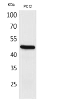
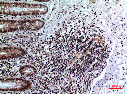
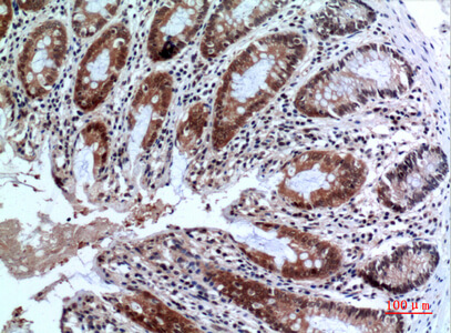
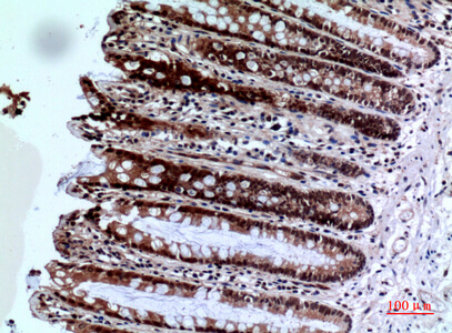
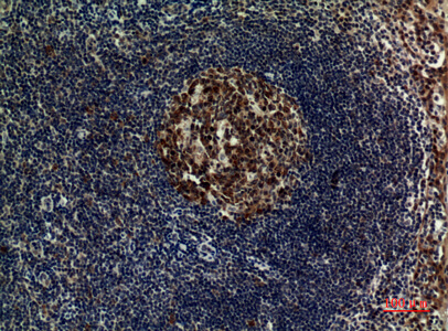
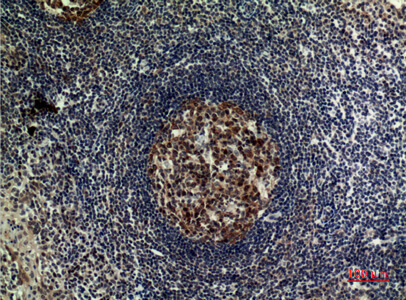
Imagine a single transcription factor that dictates the fate of our immune system—determining whether it mounts a fierce attack against pathogens and tumors or maintains peaceful tolerance to our own tissues. This molecular guardian is FOXP3, the definitive marker and functional master regulator of regulatory T cells (Tregs), a specialized lymphocyte population essential for preventing autoimmune diseases, suppressing excessive inflammation, and modulating anti-tumor immunity. The discovery of FOXP3 revolutionized immunology by providing a genetic and protein signature for Tregs, cells that constitute approximately 5–10% of peripheral CD4+ T cells and are indispensable for maintaining immune homeostasis . Mutations in the FOXP3 gene cause a devastating human autoimmune syndrome called IPEX (Immunodysregulation, Polyendocrinopathy, Enteropathy, X-linked), characterized by rampant multi-organ autoimmunity, highlighting its non-redundant role in immune suppression . Consequently, precise detection and quantification of FOXP3 protein is not merely a technical assay—it is a fundamental tool for dissecting immune tolerance mechanisms, profiling tumor microenvironments, evaluating immunotherapy efficacy, and diagnosing lymphoid malignancies. However, detecting FOXP3 presents unique challenges: it is an intranuclear protein expressed at relatively low levels, requires specific fixation and permeabilization protocols for flow cytometry, and exists in multiple isoforms due to alternative splicing . The Abbkine FOXP3 Polyclonal Antibody (ABP0141) is engineered to overcome these hurdles, offering researchers a high-affinity, rabbit-derived polyclonal reagent validated for Western blot (WB), immunohistochemistry (IHC-P), and ELISA, with confirmed reactivity against human, mouse, and rat FOXP3 . Whether you are sorting Tregs for adoptive cell therapy, analyzing FOXP3+ tumor-infiltrating lymphocytes (TILs) in cancer biopsies, or modeling autoimmune diseases in rodents, this antibody delivers the specificity, sensitivity, and reproducibility required for robust, publication-quality data across diverse experimental platforms.
FOXP3 Biology: From Transcriptional Regulator to Clinical Biomarker
FOXP3 (Forkhead box protein P3, UniProt Q9BZS1, gene ID 50943) is a 47-kDa member of the forkhead/winged-helix (FKH) family of transcription factors, characterized by a conserved DNA-binding forkhead domain, a zinc-finger motif, and a leucine zipper domain that facilitates protein-protein interactions . Unlike many transcription factors, FOXP3 expression is largely restricted to a subset of CD4+CD25+ T cells that acquire suppressive function upon activation, although it can also be transiently expressed in activated conventional T cells without conferring regulatory activity . FOXP3 functions as both a transcriptional activator and repressor: it upregulates Treg-associated genes like CTLA-4, CD25, and GITR while repressing pro-inflammatory cytokines such as IL-2 and IFN-γ . Its activity is modulated by post-translational modifications including acetylation, phosphorylation, and ubiquitination, which influence protein stability, nuclear localization, and transcriptional activity . Clinically, FOXP3 serves as a critical biomarker: high levels of FOXP3+ Tregs in tumor microenvironments correlate with poor prognosis in cancers like ovarian, breast, and hepatocellular carcinoma by suppressing anti-tumor immunity . Conversely, FOXP3 mutations or deficient Treg function underlie autoimmune conditions like type 1 diabetes, rheumatoid arthritis, and inflammatory bowel disease . In diagnostic pathology, FOXP3 immunohistochemistry helps distinguish angioimmunoblastic T-cell lymphoma (AITL) and other T-cell lymphomas from reactive lymphoid hyperplasia . Thus, reliable detection of FOXP3 protein is paramount for both basic research and translational applications.
Antody Characteristics: A Versatile, High-Performance Reagent for Multi-Platform Detection
The Abbkine FOXP3 Polyclonal Antibody (ABP0141) is an affinity-purified rabbit polyclonal antibody generated against a synthetic peptide corresponding to the C-terminal region of human FOXP3, ensuring recognition of key epitopes across multiple species and isoforms . Key specifications include:
• Host Species: Rabbit – provides high affinity and suitability for multiplex staining with mouse-derived primary antibodies in IHC/IF.
• Reactivity: Confirmed for human, mouse, and rat FOXP3, enabling cross-species studies in clinical samples and preclinical models .
• Applications: Validated for Western blot (WB), immunohistochemistry on paraffin-embedded sections (IHC-P), and ELISA . Optimal dilutions should be empirically determined but suggested starting points are:
◦ Western blot: 1:500 to 1:2000

◦ Immunohistochemistry (IHC-P): 1:100 to 1:300

◦ ELISA: 1:20000
• Clonality: Polyclonal – recognizes multiple epitopes on the FOXP3 protein, enhancing detection sensitivity and robustness, especially for denatured or partially degraded antigens in WB and IHC .
• Formulation: Liquid solution at 1 mg/mL in PBS (pH 7.4) containing 0.5% BSA (stabilizer), 0.02% sodium azide (preservative), and 50% glycerol for long-term storage .
• Storage: Stable for one year at –20°C from date of shipment; avoid repeated freeze-thaw cycles by aliquoting upon receipt .
• Immunogen: Synthetic peptide derived from the C-terminus of human FOXP3 – this region is less conserved across forkhead family members, reducing risk of cross-reactivity with FOXO1, FOXP1, etc. .
Five Transformative Applications in Modern Immunology and Oncology Research
Research Area Specific Investigation How ABP0141 Provides the Answer
Cancer Immunotherapy & Tumor Microenvironment Profiling FOXP3+ regulatory T cell infiltration in tumor biopsies (e.g., melanoma, NSCLC, colorectal cancer) to correlate with patient survival and response to immune checkpoint inhibitors (anti-PD-1/PD-L1). Perform IHC-P on FFPE tumor sections using ABP0141 (1:200) to quantify FOXP3+ TILs; high Treg density often associates with immune evasion and poor prognosis .
Autoimmune Disease Modeling Assessing Treg frequency and function in mouse models of type 1 diabetes (NOD mice), experimental autoimmune encephalomyelitis (EAE), or colitis. Use flow cytometry (with intracellular staining) or Western blot on lymph node/spleen lysates to measure FOXP3 expression, tracking disease progression or therapeutic intervention .
Stem Cell & Treg Engineering Validating FOXP3 induction during in vitro differentiation of induced pluripotent stem cells (iPSCs) into functional Tregs for adoptive cell therapy. Perform Western blot (1:1000) or immunofluorescence on day 7–14 differentiated cells to confirm nuclear FOXP3 protein expression alongside CD25 and CTLA-4 .
Lymphoma Diagnostics Distinguishing angioimmunoblastic T-cell lymphoma (AITL) from other peripheral T-cell lymphomas or reactive lymphoid hyperplasia by detecting aberrant FOXP3 expression in neoplastic T-follicular helper cells. IHC-P on lymph node biopsies with ABP0141 (1:150) shows strong nuclear staining in AITL tumor cells, aiding pathological diagnosis .
Mechanistic Studies of Treg Suppression Investigating FOXP3 phosphorylation/acetylation status under different cytokine milieus (TGF-β, IL-2) using immunoprecipitation (IP) followed by Western blot. Use ABP0141 for IP of endogenous FOXP3 from primary human Treg lysates, then probe with phospho-specific antibodies to map signaling pathways .
Step-by-Step Protocol for Reliable FOXP3 Detection Across Applications
① Western Blot (WB) for FOXP3
• Sample preparation: Lyse 1–5 × 10⁶ cells (e.g., human PBMCs, mouse splenocytes) in RIPA buffer with protease/phosphatase inhibitors. For nuclear extracts, use a nuclear extraction kit to enrich FOXP3.
• Gel electrophoresis: Load 20–40 µg total protein or 5–10 µg nuclear extract on a 10% SDS-PAGE gel. Include a positive control (e.g., lysate from activated human Tregs or FOXP3-transfected HEK293 cells).
• Transfer: Transfer to PVDF membrane at 100 V for 60 min in ice-cold transfer buffer.
• Blocking: Block with 5% non-fat milk in TBST for 1 h at room temperature.
• Primary antibody: Incubate with Abbkine FOXP3 antibody (ABP0141) diluted 1:1000 in blocking buffer overnight at 4°C with gentle shaking.
• Washing: Wash 3 × 5 min with TBST.
• Secondary antibody: Incubate with HRP-conjugated anti-rabbit IgG (1:5000) for 1 h at room temperature.
• Detection: Develop with ECL substrate and image. Expected band at ~47 kDa (full-length FOXP3); lower bands may represent isoforms or degradation products.
② Immunohistochemistry (IHC-P) on Formalin-Fixed Paraffin-Embedded (FFPE) Tissues
• Deparaffinization & antigen retrieval: Bake slides at 60°C for 1 h, deparaffinize in xylene, rehydrate through graded ethanol. Perform heat-induced epitope retrieval (HIER) in citrate buffer (pH 6.0) or EDTA buffer (pH 9.0) for 20 min in a pressure cooker or microwave.
• Peroxidase quenching: Block endogenous peroxidase with 3% H₂O₂ in methanol for 10 min.
• Blocking: Block non-specific binding with 5% normal goat serum for 30 min.
• Primary antibody: Apply ABP0141 at 1:200 dilution in antibody diluent and incubate overnight at 4°C in a humidified chamber.
• Detection: Use a polymer-based HRP detection system (e.g., DAB) following manufacturer’s instructions. Counterstain with hematoxylin, dehydrate, and mount.
• Interpretation: Nuclear staining in lymphocytes is specific; cytoplasmic staining may indicate non-specific binding or improper fixation.
③ Intracellular Flow Cytometry for FOXP3 in T Cells
• Cell stimulation & fixation: Stimulate PBMCs or isolated CD4+ T cells with PMA/ionomycin + protein transport inhibitor (e.g., brefeldin A) for 4–6 h. Fix with 4% paraformaldehyde (PFA) for 20 min at room temperature.
• Permeabilization: Permeabilize with ice-cold methanol or a commercial permeabilization buffer (e.g., Foxp3/Transcription Factor Staining Buffer Set) for 30 min on ice.
• Staining: Incubate with surface antibodies (anti-CD4, anti-CD25) first, then with ABP0141 (1:100–1:500) in permeabilization buffer for 30 min at 4°C, followed by fluorophore-conjugated anti-rabbit secondary.
• Acquisition: Analyze on a flow cytometer; FOXP3+ cells typically appear within the CD4+CD25high population.
④ ELISA for Quantifying Soluble FOXP3?
• Note: While ABP0141 is listed for ELISA, FOXP3 is primarily an intranuclear protein with minimal secreted forms. ELISA applications are less common and typically measure cleaved or alternative isoforms in serum/plasma, which may have limited biological relevance. For classical FOXP3 detection, WB, IHC, or flow cytometry are preferred.
Troubleshooting Guide for Optimal FOXP3 Staining
Issue Potential Cause Recommended Solution
Weak or no signal in Western blot Insufficient protein loading (FOXP3 is low-abundance); over-transfer (protein lost); inefficient antigen retrieval for nuclear protein. Load 30–50 µg of nuclear extract; optimize transfer time (60–90 min); include a positive control lysate (e.g., FOXP3-transfected cells).
High background in IHC Non-specific binding of primary or secondary antibody; incomplete blocking; endogenous peroxidase activity not fully quenched. Use species-appropriate normal serum blocking (5% goat serum for rabbit primary); extend H₂O₂ quenching to 15 min; titrate primary antibody (try 1:400–1:800).
Non-nuclear (cytoplasmic) staining in IHC/IF Over-fixation (formalin cross-linking masks nuclear epitopes); inadequate antigen retrieval; antibody cross-reactivity. Optimize antigen retrieval time/temperature (try 20 min pressure cooking in pH 9.0 EDTA buffer); use monoclonal anti-FOXP3 clone 236A/E7 for comparison.
Poor resolution in flow cytometry Inadequate permeabilization (methanol may be too harsh for some epitopes); antibody concentration too high causing aggregation. Test commercial Foxp3 staining buffers (e.g., eBioscience); titrate ABP0141 from 1:100 to 1:1000; include fluorescence-minus-one (FMO) controls.
Multiple bands in Western blot FOXP3 isoforms (e.g., ~41 kDa splice variant); proteolytic degradation; non-specific binding. Use fresh protease inhibitors; run a high-percentage gel (12%) for better resolution; confirm identity with FOXP3 siRNA knockdown.
Inconsistent staining between experiments Lot-to-lot variability (though minimal with affinity purification); differences in fixation protocols; antibody storage degradation. Aliquot antibody upon arrival; avoid repeated freeze-thaw; standardize fixation time across samples; record antibody lot number for reproducibility.
Benchmarking: Polyclonal vs. Monoclonal FOXP3 Antibodies
Parameter Abbkine Polyclonal (ABP0141) Monoclonal (e.g., clone 236A/E7)
Epitope Recognition Multiple epitopes (C-terminal peptide), may detect isoforms and denatured forms. Single epitope (often N-terminal), may miss certain isoforms or modified forms.
Sensitivity High – multiple epitopes increase chance of binding, advantageous for low-abundance targets. Moderate – depends on epitope accessibility; may require stronger antigen retrieval.
Specificity Potential cross-reactivity with other forkhead proteins if epitope is conserved; requires careful validation. High – single epitope reduces off-target binding; preferred for diagnostic IHC.
Application Flexibility Excellent for WB, IHC-P, ELISA – robust across platforms due to epitope diversity. Best for flow cytometry and IHC where consistency and specificity are critical.
Cost More economical per microgram. Typically more expensive due to hybridoma maintenance and purification.
Recommended Use Research applications where detection of multiple isoforms or denatured protein is needed; multiplex IHC with mouse primaries. Clinical diagnostics, flow cytometry panels requiring high specificity and reproducibility.
The Abbkine polyclonal antibody offers a cost-effective, sensitive option for researchers exploring FOXP3 across multiple techniques, especially when studying protein isoforms or working with denatured samples from Western blot.
Best Practices for Publication-Quality FOXP3 Data
Practice Rationale & Implementation
Validate antibody specificity in your system Perform siRNA/shRNA knockdown of FOXP3 in a relevant cell line (e.g., Jurkat T cells) followed by Western blot to confirm band disappearance at ~47 kDa.
Use appropriate positive and negative controls For IHC/flow, include human tonsil or thymus (rich in Tregs) as positive control; FOXP3-negative cell lines (e.g., HeLa) as negative control.
Optimize antigen retrieval for IHC Test both citrate (pH 6.0) and EDTA (pH 9.0) buffers with heat retrieval; FOXP3 often requires high-pH EDTA buffer for optimal nuclear exposure.
Combine with lineage markers for flow cytometry Always co-stain with CD4, CD25, and CD127 to accurately identify CD4+CD25+CD127lowFOXP3+ natural Tregs; exclude dead cells with viability dye.
Quantify IHC results objectively Use digital pathology software (e.g., QuPath, HALO) to count FOXP3+ nuclei per mm² or calculate percentage of FOXP3+ cells among CD3+ T cells.
Document antibody dilution and lot number Record exact dilution, incubation time/temperature, and lot #ABP0141-XXX in methods; essential for reproducibility and manuscript submission.
Store antibody properly Aliquot upon receipt; store at –20°C; avoid repeated freeze-thaw (>3 cycles); glycerol in formulation prevents freezing at –20°C, but long-term storage at –80°C is recommended.
From Basic Research to Clinical Translation: Key Insights Enabled by FOXP3 Detection
① Unraveling Treg biology in autoimmunity
Using ABP0141 for Western blot and flow cytometry, researchers have demonstrated that FOXP3 expression is unstable in inflammatory environments (e.g., high IL-6), leading to Treg plasticity and loss of suppressive function—a mechanism implicated in rheumatoid arthritis and type 1 diabetes flare-ups .
② Prognostic biomarker in oncology
IHC with ABP0141 on tumor microarrays has revealed that high FOXP3+ Treg density in the tumor stroma correlates with poor clinical outcome in ovarian, breast, and gastric cancers, supporting Tregs as a therapeutic target to enhance checkpoint inhibitor efficacy .
③ Monitoring immunotherapy responses
In patients receiving anti-CTLA-4 (ipilimumab) or anti-PD-1 therapy, tracking FOXP3+ Treg frequency in peripheral blood via flow cytometry can serve as a dynamic pharmacodynamic biomarker, with early decreases often associated with clinical response .
④ Engineering CAR-Tregs for tolerance induction
FOXP3 staining is critical for quality control during manufacturing of chimeric antigen receptor (CAR)-Tregs for treating graft-versus-host disease (GVHD) and organ transplant rejection, ensuring engineered cells maintain stable FOXP3 expression .
⑤ Diagnostic aid in hematopathology
FOXP3 IHC helps differentiate angioimmunoblastic T-cell lymphoma (AITL) from other peripheral T-cell lymphomas, as neoplastic T-follicular helper cells in AITL frequently express FOXP3, a feature used in the WHO classification .
A Ready-to-Use Methods Section for Your Manuscript
FOXP3 protein expression was analyzed by Western blot using the FOXP3 Polyclonal Antibody (Abbkine, ABP0141). Briefly, nuclear extracts were prepared from 2 × 10⁶ cells using a nuclear extraction kit (NE-PER, Thermo Scientific). Proteins (30 µg per lane) were separated on a 10% SDS-PAGE gel and transferred to PVDF membranes. Membranes were blocked with 5% non-fat milk in TBST for 1 h, then incubated overnight at 4°C with rabbit anti-FOXP3 antibody (ABP0141, 1:1000 dilution). After washing, membranes were incubated with HRP-conjugated goat anti-rabbit IgG (1:5000) for 1 h at room temperature. Signals were detected using enhanced chemiluminescence (ECL) and imaged on a ChemiDoc system (Bio-Rad). β-actin or lamin B1 served as loading controls. For immunohistochemistry, formalin-fixed, paraffin-embedded tissue sections were deparaffinized, subjected to heat-induced epitope retrieval in EDTA buffer (pH 9.0), and stained with ABP0141 (1:200) using a polymer-HRP detection system (DAB). FOXP3+ cells were quantified as the number of positive nuclei per high-power field (HPF) using ImageJ software.
Why the Abbkine FOXP3 Polyclonal Antibody (ABP0141) Is a Cornerstone Reagent for Immunology Research
① Broad species reactivity – validated for human, mouse, and rat FOXP3, enabling translational studies from preclinical models to clinical samples .
② Multi-platform versatility – optimized for Western blot, IHC-P, and ELISA, providing a single reagent for diverse experimental needs, from protein quantification to tissue localization .
③ High sensitivity and affinity – affinity-purified polyclonal antibody offers superior detection of low-abundance nuclear FOXP3, even in partially degraded or denatured samples .
④ Cost-effective without compromising performance – compared to many monoclonal antibodies, ABP0141 provides excellent value per microgram while maintaining high batch-to-batch consistency .
⑤ Robust technical support – each lot is quality-controlled for specificity and reactivity, and Abbkine provides detailed protocols and troubleshooting assistance to ensure experimental success.
⑥ Critical for cutting-edge applications – essential for Treg isolation, tumor immunology profiling, autoimmune disease modeling, and immunotherapy monitoring, supporting research published in high-impact journals like Nature Immunology, Cancer Cell, and Journal of Autoimmunity.
Ready to decode the secrets of immune tolerance? The Abbkine FOXP3 Polyclonal Antibody (ABP0141) delivers unmatched versatility, sensitivity, and reliability—whether you're mapping Treg infiltration in tumors, modeling autoimmune disorders, or engineering next-generation regulatory cell therapies. With cross-species reactivity, multi-application validation, and robust performance, it's the definitive tool for illuminating the FOXP3 pathway.
🔗 Product reference: ABP0141 (Abbkine) – https://www.abbkine.com/product/foxp3-polyclonal-antibody-abp0141/
(For research use only. Not for diagnostic or therapeutic procedures. Store at –20°C protected from light; stable for 12 months from date of shipment.)





