


Decoding the Cellular Stress Nexus: The Abbkine JNK1/2/3 (phospho Thr183/Y185) Polyclonal Antibody for Precision Signal Transduction Research
When cells face environmental assaults—be it oxidative stress, inflammatory cytokines, or DNA damage—a rapid and precise molecular decision-making cascade is triggered to determine fate: adapt, repair, or undergo programmed death. At the heart of this critical response lies the c-Jun N-terminal Kinase (JNK) pathway, a pivotal branch of the Mitogen-Activated Protein Kinase (MAPK) signaling network . The activation of JNK isoforms (JNK1, JNK2, JNK3) is a tightly regulated event, culminating in the dual phosphorylation of threonine and tyrosine residues within the Thr-Pro-Tyr motif (Thr183 and Tyr185) . This specific phosphorylation event is the definitive molecular signature of JNK activation, transforming it into a potent kinase that phosphorylates key substrates like c-Jun, ATF2, and p53, thereby influencing gene expression, proliferation, differentiation, and apoptosis . For researchers dissecting the mechanisms of cancer development, neurodegenerative diseases, inflammatory disorders, metabolic syndrome, and response to therapeutics, the ability to accurately detect and quantify this activation event is non-negotiable. The JNK1/2/3 (phospho Thr183/Y185) Polyclonal Antibody (ABP50351) from Abbkine is a meticulously validated, phospho-specific tool designed to illuminate this crucial signaling node. It enables the specific, sensitive, and reliable detection of dually phosphorylated, activated JNK1, JNK2, and JNK3 across a wide range of applications, including Western Blot (WB), Immunohistochemistry (IHC), Immunofluorescence (IF), and Immunoprecipitation (IP) . Whether you are mapping stress kinase activation in tumor microenvironments, investigating JNK's role in neuronal apoptosis in Alzheimer's models, studying its contribution to insulin resistance, or screening for novel kinase inhibitors, this antibody provides the specificity and reproducibility required to generate conclusive, publication-ready data.
JNK Phosphorylation: The Master Switch in Cellular Stress Signaling
The JNK pathway is a central mediator of cellular responses to a diverse array of extracellular stresses and intracellular cues . Activation occurs through a sequential kinase cascade: MAPK kinase kinases (MAP3Ks, like ASK1, MLKs) phosphorylate and activate MAPK kinases (MAP2Ks, MKK4/7), which in turn dually phosphorylate JNK on Thr183 and Tyr185 . This phospho-Thr183/Tyr185 epitope is the universal marker for active JNK across all its isoforms. Once activated, phospho-JNK translocates to the nucleus where it phosphorylates transcription factors, primarily components of the AP-1 complex like c-Jun, enhancing their transcriptional activity and driving the expression of genes involved in inflammation, survival, and apoptosis . The biological outcomes are context-dependent: sustained JNK activation often promotes apoptotic cell death, as seen in neurons exposed to amyloid-beta or during hepatic injury, while transient activation may support cell proliferation and survival, contributing to tumorigenesis in certain cancers . Its involvement is implicated in a vast spectrum of pathologies, including cancer (e.g., liver, breast, lung), neurodegenerative diseases (Alzheimer's, Parkinson's), inflammatory conditions (rheumatoid arthritis, asthma), and metabolic disorders (type 2 diabetes, non-alcoholic fatty liver disease) . Therefore, monitoring JNK phosphorylation status is not merely an observation of a biochemical event; it is a direct readout of a cell's stress signaling status and a key indicator of pathological processes.
Antody Specificity and Validation: A Key to Reliable Detection
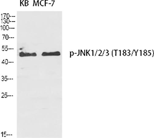
The Abbkine JNK1/2/3 (phospho Thr183/Y185) Polyclonal Antibody (ABP50351) is generated using a synthetic phosphopeptide corresponding to the conserved activation loop region surrounding dually phosphorylated Thr183 and Tyr185. This design ensures high affinity and specificity for the phosphorylated form of JNK. Rigorous validation confirms that it recognizes phospho-JNK1 (p46/p54), phospho-JNK2 (p46/p54), and phospho-JNK3 (p46/p54), with minimal to no cross-reactivity with non-phosphorylated JNK or other phosphorylated MAPK family members like ERK or p38 . This specificity is paramount, as it allows researchers to distinguish the active, signaling-competent pool of JNK from the total pool of the protein, a critical distinction for mechanistic studies. The antibody's performance is consistently demonstrated in multiple applications, providing versatility for different experimental needs.
Key Features and Application Performance of ABP50351
• High Specificity for Phospho-JNK: Specifically detects JNK1, JNK2, and JNK3 only when phosphorylated at Thr183 and Tyr185. Does not cross-react with corresponding non-phosphorylated proteins or other phospho-MAPKs .
• Broad Reactivity and Isoform Coverage: Recognizes all major isoforms of phosphorylated JNK (JNK1, JNK2, JNK3) across multiple species, including human, mouse, and rat, making it suitable for a wide range of preclinical models .
• Versatile Application Suitability: This polyclonal antibody is rigorously validated for use in multiple key techniques:
◦ Western Blotting (WB): Delivers clear, specific bands at the expected molecular weights (~46 kDa and ~54 kDa) for phospho-JNK from whole cell lysates, tissue homogenates, or subcellular fractions. Ideal for quantifying activation dynamics in response to stimuli (e.g., anisomycin, UV radiation, TNF-α) or drug treatments .
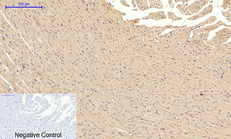
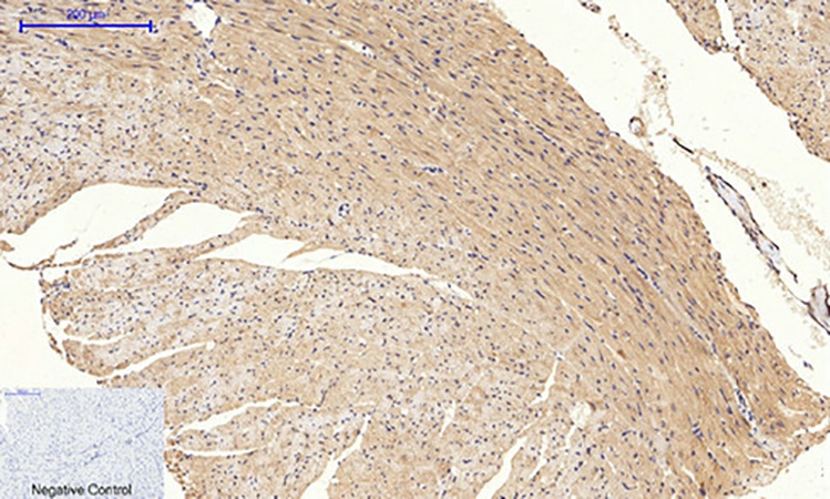
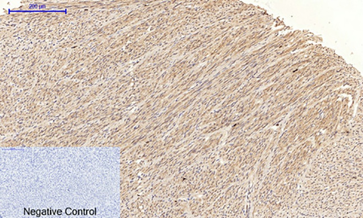
◦ Immunohistochemistry (IHC) & Immunofluorescence (IF): Enables the spatial visualization of activated JNK within fixed tissues or cells. This is crucial for identifying which cell types within a complex tissue (e.g., tumor stroma vs. cancer cells, neurons vs. glia) exhibit active JNK signaling, providing critical pathological insights .
◦ Immunoprecipitation (IP): Can be used to pull down active, phosphorylated JNK from complex lysates for downstream analysis of interacting partners or kinase activity assays .
• Superior Sensitivity and Signal-to-Noise Ratio: Optimized formulations and validation ensure strong, specific signals with low background across recommended applications, allowing for the detection of phospho-JNK even in samples with lower expression levels.
• Consistent Lot-to-Lot Performance: Manufactured under stringent quality control to ensure high batch-to-batch consistency, providing reliable and reproducible results for long-term studies.
Critical Research Applications Enabled by the Phospho-JNK Antibody
- Cancer Research: Investigate the role of sustained JNK activation in tumor cell survival, proliferation, and chemo-resistance. Correlate phospho-JNK levels with tumor grade, stage, and patient prognosis in tissue microarray (TMA) studies using IHC. Study crosstalk between oncogenic signaling and stress pathways .
- Neuroscience and Neurodegeneration: Map JNK activation in models of Alzheimer's disease, Parkinson's disease, and stroke. Determine its contribution to neuronal apoptosis, synaptic dysfunction, and neuroinflammation using IF on brain sections or WB of neuronal lysates .
- Inflammation and Immunology: Analyze JNK pathway activation in immune cells (e.g., T cells, macrophages) in response to pro-inflammatory cytokines (TNF-α, IL-1β). Study its role in autoimmune diseases and acute inflammatory responses .
- Metabolic Disease Studies: Examine the link between metabolic stress (e.g., lipotoxicity, glucotoxicity), JNK activation, and insulin resistance in liver, muscle, or adipose tissue models. A key tool for diabetes and NAFLD/NASH research .
- Drug Discovery and Toxicology: Utilize phospho-JNK detection as a pharmacodynamic biomarker to assess the efficacy of novel JNK inhibitors or other therapeutics targeting upstream kinases. Monitor pathway modulation in cell-based assays and in vivo models .
- Basic Cell Signaling: Elucidate the temporal dynamics of JNK activation following various cellular stresses (UV, osmotic shock, DNA damaging agents) and its integration with other signaling networks like PI3K/Akt and NF-κB .
Streamlined Workflow for Western Blot Analysis (Example)
Step 1: Sample Preparation and Electrophoresis. Lyse cells or tissues in a suitable RIPA buffer supplemented with phosphatase inhibitors (essential to preserve phosphorylation) and protease inhibitors. Determine protein concentration, prepare samples with Laemmli buffer, and denature. Load equal amounts of protein onto an SDS-PAGE gel (e.g., 10%) and separate by electrophoresis.
Step 2: Transfer and Blocking. Transfer proteins from the gel to a PVDF or nitrocellulose membrane. Block the membrane with 5% non-fat dry milk or BSA in TBST for 1 hour at room temperature to prevent non-specific binding.
Step 3: Primary Antibody Incubation. Dilute the Abbkine JNK1/2/3 (phospho Thr183/Y185) Polyclonal Antibody (ABP50351) in the recommended antibody dilution buffer (e.g., 5% BSA in TBST). A typical starting dilution for WB is 1:1000. Incubate the membrane with the primary antibody solution overnight at 4°C with gentle agitation.
Step 4: Washing and Secondary Antibody Incubation. Wash the membrane 3-4 times with TBST for 5-10 minutes each. Incubate with an appropriate HRP-conjugated secondary antibody (e.g., goat anti-rabbit IgG-HRP) diluted in blocking buffer for 1 hour at room temperature. Wash again thoroughly.
Step 5: Detection and Analysis. Develop the signal using a sensitive chemiluminescent substrate and image with a digital imager. Expected bands are at ~46 kDa and ~54 kDa. For loading control, strip and re-probe the membrane with an antibody against total JNK or β-actin.
Why Choose the Abbkine Phospho-JNK (Thr183/Y185) Polyclonal Antibody?
• Confidence in Specificity: The phospho-specific design ensures you are detecting the biologically active form of JNK, providing clear mechanistic insights that total JNK antibodies cannot offer.
• Versatility for Multi-Modal Analysis: Validated for WB, IHC, IF, and IP, this single antibody empowers you to study JNK activation from the level of whole tissue architecture down to specific protein interactions, maximizing the return on your research investment.
• Reliability for Demanding Applications: The consistent performance and low background ensure high-quality, reproducible data in challenging applications like immunohistochemistry on archival FFPE tissues, where signal clarity is paramount.
• A Catalyst for Discovery: By providing a reliable tool to monitor this central stress signaling node, this antibody accelerates research into fundamental biology and disease mechanisms, from cancer therapy resistance to neuroprotective strategies.
Illuminate the path of cellular decision-making. The Abbkine JNK1/2/3 (phospho Thr183/Y185) Polyclonal Antibody (ABP50351) is an essential, high-quality reagent for any laboratory focused on MAPK signaling, stress response, and their roles in human disease.
Product Reference: ABP50351 – JNK1/2/3 (phospho Thr183/Y185) Polyclonal Antibody
Learn more and order: https://www.abbkine.com/product/jnk1-2-3-phospho-thr183-y185-polyclonal-antibody-abp50351/


