


The Master Switch of Proliferation and Survival: Illuminating ERK1/2 Activation with Abbkine's Phospho-Thr202/Y204 Specific Antibody
At the core of countless cellular decisions—from growth and differentiation to adaptation and survival—lies a highly conserved signaling cascade whose activity is governed by a single, definitive molecular event. The Extracellular Signal-Regulated Kinases 1 and 2 (ERK1/2) pathway stands as a central conductor of mitogenic and developmental signals, translating extracellular cues into precise intracellular responses . Its activation, marked by the dual phosphorylation of threonine and tyrosine residues within the Thr-Glu-Tyr motif (Thr202 and Tyr204 in ERK1, Thr185 and Tyr187 in ERK2), serves as the critical on-switch that unleashes its kinase activity . This phosphorylation event, catalyzed by the upstream kinase MEK, triggers ERK1/2 dimerization, cytoplasmic-to-nuclear translocation, and the phosphorylation of hundreds of substrates, including transcription factors like Elk-1 and c-Fos, thereby orchestrating gene expression programs . For researchers across cancer biology, neuroscience, immunology, and stem cell research, accurately detecting this phospho-epitope is not just a technical step; it is a direct readout of pathway activity, a biomarker for oncogenic signaling, and a key endpoint for therapeutic intervention . The ERK 1/2 (phospho Thr202/Y204) Polyclonal Antibody (ABP50531) from Abbkine is a rigorously validated, phospho-specific reagent designed to capture this pivotal moment with high fidelity. It enables the specific, sensitive, and consistent detection of activated ERK1 and ERK2 across a multitude of applications, including Western Blot (WB), Immunohistochemistry (IHC), Immunofluorescence (IF), and ELISA . Whether you are tracking oncogenic RAS/RAF/MEK/ERK pathway activation in tumor samples, mapping neuronal ERK signaling in learning and memory models, studying T-cell receptor engagement, or validating the efficacy of novel MEK inhibitors, this antibody provides the critical specificity needed to generate definitive, publication-quality data.
ERK1/2 Phosphorylation: The Definitive Signal for Cellular Growth and Fate
The ERK1/2 cascade is arguably the most studied MAPK pathway, acting as a primary signaling hub for receptor tyrosine kinases (RTKs), G-protein coupled receptors (GPCRs), and integrins . Upon ligand binding and receptor activation, a sequential kinase relay (RAS → RAF → MEK) culminates in MEK1/2 phosphorylating ERK1/2 on the conserved Thr202 and Tyr204 residues . This dual phosphorylation induces a conformational change that fully activates ERK1/2 kinase function . Activated phospho-ERK1/2 then phosphorylates a vast array of cytoplasmic and nuclear targets, regulating immediate early gene expression, protein synthesis, cell cycle progression, and cytoskeletal dynamics . The duration and amplitude of ERK1/2 phosphorylation encode specific biological outcomes: transient activation often promotes proliferation and differentiation, while sustained activation can lead to cell cycle arrest, senescence, or even apoptosis in certain contexts . Dysregulation of this pathway is a hallmark of human disease, most notably in cancer, where hyperactivation via mutations in upstream components (e.g., BRAF V600E, KRAS G12D) drives uncontrolled growth and survival in melanomas, lung adenocarcinomas, and colorectal cancers . It also plays crucial roles in neuronal plasticity, cardiac hypertrophy, and immune cell activation . Therefore, monitoring the phosphorylation status of ERK1/2 at Thr202/Tyr204 is essential for understanding both normal physiology and the pathogenesis of numerous disorders.
Antibody Design and Specificity: A Key to Accurate Pathway Interrogation
The Abbkine ERK 1/2 (phospho Thr202/Y204) Polyclonal Antibody (ABP50531) is generated using a synthetic phosphopeptide corresponding to the conserved activation loop region surrounding dually phosphorylated Thr202 and Tyr204 of human ERK1. This immunogen design ensures high affinity and exceptional specificity for the phosphorylated, active form of ERK1 and ERK2 . Extensive validation confirms that it recognizes phospho-ERK1 (p44) and phospho-ERK2 (p42) with minimal cross-reactivity to non-phosphorylated ERK or other phosphorylated MAPKs like JNK or p38 . This specificity is paramount, as it allows researchers to clearly distinguish the active, signaling-competent pool of ERK from the total cellular pool—a distinction that is fundamental for mechanistic studies and pharmacodynamic assessments. The antibody's robust performance across multiple platforms makes it a versatile cornerstone for signal transduction research.
Key Features and Validated Application Performance
• High Specificity for Phospho-ERK1/2: Specifically detects ERK1 and ERK2 only when phosphorylated at Thr202 and Tyr204 (Thr185/Tyr187 for ERK2). Shows negligible reactivity with the non-phosphorylated proteins, ensuring a clean signal for pathway activation .
• Broad Species Reactivity: Effectively recognizes phosphorylated ERK1/2 from human, mouse, and rat samples, making it suitable for a wide range of in vitro and in vivo preclinical models .
• Versatile Multiplex Application Suitability: This polyclonal antibody is rigorously validated for use in key experimental techniques:
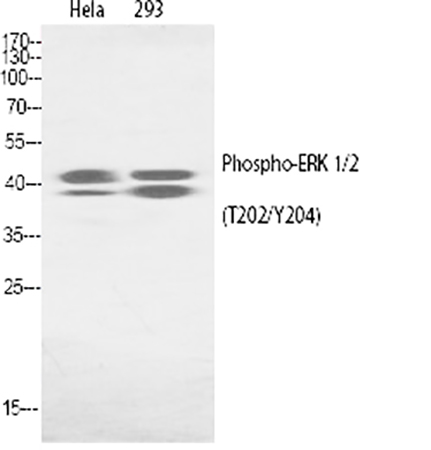
◦ Western Blotting (WB): Produces clear, specific bands at the expected molecular weights (~44 kDa for ERK1/p44 and ~42 kDa for ERK2/p42) from whole cell lysates, tissue homogenates, or subcellular fractions. Ideal for kinetic studies of pathway activation in response to growth factors (EGF, FGF), serum, or pharmacological inhibitors .
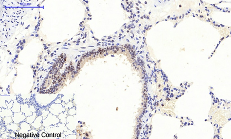
◦ Immunohistochemistry (IHC) & Immunofluorescence (IF): Enables precise spatial visualization of activated ERK1/2 within the architecture of fixed tissues or cultured cells. This is critical for identifying which cell populations within a tumor, brain region, or developing tissue exhibit active MAPK signaling .
◦ Enzyme-Linked Immunosorbent Assay (ELISA): Can be configured for quantitative measurement of phospho-ERK1/2 levels in cell lysates, providing a high-throughput method for screening compound libraries or analyzing clinical samples .
• Optimized for Sensitivity and Low Background: The antibody is supplied in a formulation that delivers strong, specific signals with minimal background noise across its recommended applications, facilitating the detection of phospho-ERK even in samples with modest activation levels.
• Consistent and Reliable Performance: Manufactured under stringent quality control protocols to ensure high batch-to-batch consistency, providing dependable results for longitudinal and multi-center studies.
Pivotal Research Applications Enabled by the Phospho-ERK1/2 Antibody
- Cancer Research and Biomarker Discovery: Investigate the activation status of the RAS/RAF/MEK/ERK pathway in patient-derived tumor tissues using IHC. Correlate phospho-ERK1/2 levels with tumor grade, stage, mutational status (e.g., BRAF, KRAS), and patient response to targeted therapies like MEK inhibitors .
- Drug Discovery and Target Validation: Utilize WB or ELISA to monitor phospho-ERK1/2 as a primary pharmacodynamic biomarker in cell-based and in vivo models treated with novel RTK inhibitors, RAF inhibitors, or MEK inhibitors, accelerating the preclinical development of targeted oncology drugs .
- Neuroscience and Synaptic Plasticity: Map ERK activation in models of learning and memory, such as long-term potentiation (LTP), and in neurological disorders. Study its role in neuronal survival, differentiation, and response to neurotrophic factors .
- Developmental Biology and Stem Cell Research: Analyze ERK1/2 signaling dynamics during embryonic development, tissue morphogenesis, and stem cell differentiation. Determine how growth factor gradients are translated into spatial patterns of pathway activation .
- Immunology and Inflammation: Assess ERK1/2 phosphorylation in immune cells (e.g., T cells, B cells, macrophages) following antigen receptor engagement or cytokine stimulation to understand its role in immune cell activation, proliferation, and cytokine production .
- Cardiovascular Research: Examine the contribution of ERK1/2 activation to cardiac myocyte hypertrophy, survival, and remodeling in response to pressure overload or neurohormonal stimuli.
Recommended Protocol for Western Blot Analysis
Step 1: Sample Preparation with Phosphatase Inhibition. Lyse cells or tissues in a suitable RIPA or NP-40 buffer supplemented with both protease and phosphatase inhibitors. This step is critical to preserve the labile phosphorylation state of ERK1/2. Determine protein concentration, mix samples with reducing Laemmli buffer, denature by heating, and load equal amounts (typically 20-40 µg) onto an SDS-PAGE gel (10-12%).
Step 2: Electrophoresis and Transfer. Separate proteins by standard SDS-PAGE. Transfer proteins from the gel to a PVDF or nitrocellulose membrane using a wet or semi-dry transfer system.
Step 3: Blocking and Primary Antibody Incubation. Block the membrane with 5% non-fat dry milk or 3-5% BSA in TBST for 1 hour at room temperature. Incubate the membrane with the Abbkine Phospho-ERK1/2 (Thr202/Y204) Antibody (ABP50531), typically diluted at 1:1000 in 5% BSA/TBST, overnight at 4°C with gentle agitation.
Step 4: Washing and Secondary Detection. Wash the membrane 3-4 times for 5-10 minutes each with TBST. Incubate with an HRP-conjugated anti-rabbit secondary antibody (diluted 1:5000-1:10000 in blocking buffer) for 1 hour at room temperature. Wash thoroughly again.
Step 5: Signal Development and Normalization. Develop the signal using a sensitive chemiluminescent substrate and capture the image. The expected bands are at ~44 kDa (phospho-ERK1) and ~42 kDa (phospho-ERK2). To control for loading, strip the membrane (if necessary) and re-probe with an antibody against total ERK1/2 or a housekeeping protein like β-actin or GAPDH.
Why the Abbkine Phospho-ERK1/2 (Thr202/Y204) Antibody is a Laboratory Essential
• Unambiguous Pathway Readout: The phospho-specificity provides a direct and accurate measure of ERK1/2 pathway activation, eliminating ambiguity and enabling clear interpretation of signaling events in complex biological systems.
• Flexibility Across Experimental Platforms: Validated for WB, IHC, IF, and ELISA, this single antibody empowers researchers to interrogate ERK activation at multiple levels—from molecular weight confirmation in lysates to spatial localization in tissue context—maximizing utility and return on investment.
• Trusted Reproducibility: The consistent lot-to-lot performance and low background ensure reliable, high-quality data that forms a solid foundation for hypothesis testing and publication, saving time and resources on optimization.
• A Catalyst for Translational Insights: By providing a robust tool to monitor this central growth and survival pathway, this antibody accelerates research from fundamental mechanism discovery to the evaluation of therapeutic strategies in cancer and beyond.
Illuminate the decisive moments in cellular signaling. The Abbkine ERK 1/2 (phospho Thr202/Y204) Polyclonal Antibody (ABP50531) is an indispensable, high-performance tool for any laboratory dedicated to unraveling the complexities of MAPK signaling in health and disease.
Product Reference: ABP50531 – ERK 1/2 (phospho Thr202/Y204) Polyclonal Antibody
Learn more and order: http://www.abbkine.com/product/erk-1-2-phospho-thr202-y204-polyclonal-antibody-abp50531/


