


Abbkine takes you to understand apoptosis detection!
Apoptosis:Apoptosis refers to the autonomous and orderly death of cells controlled by genes in order to maintain the stability of the internal environment. Apoptosis is not a passive process, but an active process, which involves the activation, expression and regulation of a series of genes. It is not a phenomenon of self-injury under pathological conditions, but a death process that strives for better adaptation to the living environment.
Common methods for detection of apoptosis:
| Detection method | example |
| Annexin V detection | Annexin V/PI double staining |
| Mitochondrial membrane potential-dependent dye | JC-1 |
| DNA consolidation and fragmentation | TUNEL detection |
| Active caspase detection | Caspase 3 |
| Cytochrome C release | cytochrome c |
Among them, the most commonly used methods are:
1.Detection of Annexin V: Annexin V/PI double staining
In normal cells, phosphatidylserine (PS) on the cell membrane is located on the cytoplasmic side. In the mid-apoptotic phase, PS is turned over. Annexin V is a Ca2+-dependent phospholipid binding protein with high affinity to PS, so the fluorescently labeled Annexin V can be used to detect PS eversion. At the end of apoptosis, the cell membrane structure is damaged, and nucleic acid dyes (such as PI, 7-AAD, etc.) that are impermeable to the membrane can also enter the cell to generate signals.
Annexin V/PI double staining method for detection of apoptosis experimental procedures and common problems analysis:
a.The problem of cell state represented by each quadrant
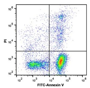
- Q1:(AnnexinV-dye) -/PI+, the cells in this area are necrotic cells. There may also be a small number of late apoptotic cells, even mechanically damaged cells. The more Annexin V-/PI+, the more unreliable the experimental results. It is recommended to control the ratio of cells in this quadrant as much as possible in the experiment;
- Q2:(AnnexinV+ dye) +/PI+, the cells in this area are late apoptotic cells;
- Q3:(AnnexinV-dye)+/PI-, the cells in this area are early apoptotic cells;
- Q4:(AnnexinV-dye)-/PI-, the cells in this area are living cells.
b.False positive
For adherent cells, digestion is the most critical step. Trypsin without EDTA should be used for digestion, because the combination of Annexin V and PS requires Ca2+, and EDTA is a Ca2+ chelating agent. The use of pancreatin containing EDTA will cause False negatives. Secondly, the digestion time should not be too long, and the cells should be gently pipetted, because excessive digestion of pancreatin and mechanical damage will cause cell membrane damage, resulting in false positives.
Nerve cell membranes are easy to damage eversion and cause false positives. Therefore, the Annexin V/PI double staining method for flow cytometry detection of apoptosis is not suitable for nerve cells.
c.Can the cells be fixed before or after staining?
Fixation will destroy the integrity of the cell, allow PI to enter the cell through the cell membrane, and increase the percentage of apoptosis of the cell. Therefore, the cells before and after staining can not be fixed, and the cells after staining need to be detected by the upstream flow method within 1 hour.
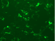
2.Tunel method detection
In cell apoptosis, chromosomal DNA double-strand breaks or single-strand breaks produce a large number of sticky 3'-OH ends. Under the action of deoxyribonucleotide terminal transferase (TdT), deoxyuridine triphosphate nucleoside The derivative formed by acid (dUTP) and fluorescein is labeled to the 3'-end of DNA, so that apoptotic cells can be detected. This type of method is called deoxyribonucleotide terminal transferase-mediated nick end labeling. Since normal or proliferating cells have almost no DNA breaks, there is no 3'-OH formation, and they are rarely stained. As a result, TUNEL has become the most common method for detecting DNA fragmentation (apoptosis).
Common problems and solutions of apoptosis detected by TUNEL method:
a.How to set the positive group, negative group and experimental group
Positive group: treated with DNase enzyme to induce cell apoptosis and expose the 3'-OH end. Just do one for each experiment;
Negative group: No TdT enzyme is added, and the rest of the operations are the same as the experimental group
Experimental group: Except for the positive group and the negative group, all samples are the experimental group.
b.Weak or no fluorescence signal
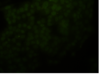
Improper sample handling
- The sample section is too thick. It is recommended to cut the slices a bit thinner to facilitate staining;
- Inadequate dewaxing and hydration. It is recommended to first dewax at 60 ºC for 20 minutes, and then use xylene twice for 5-10 minutes; and for hydration, use gradient ethanol to immerse from high to low concentration;
- The incubation time of Proteinase K is too short. Optimize the incubation time of Proteinase K, the usual time is 10-30 min. Incubate for 10 min for slices of about 4 μm, and for 30 min for slices of about 30 μm;
- The concentration of Proteinase K is too low. It is recommended that the concentration of the general working solution of proteinase K is 20 μg/mL;
- Sections stored at -20 ºC for a long time are not fresh, reducing staining efficiency. It is recommended to use fresh slices.
Improper dyeing operation
- The dyeing time is too short. It is recommended to incubate at 37 ºC for 60 minutes. Depending on the degree of apoptosis damage, it can be as long as 2 hours, but it should be combined with background coloring;
- The concentration of TdT enzyme or luciferin/enzyme-labeled dUTP is too low. It is recommended to appropriately increase the concentration of TdT enzyme or luciferin/enzyme-labeled dUTP;
- Inactivation of TdT enzyme. It is recommended that the TUNEL reaction solution be prepared immediately before use and stored on ice for a short time. It is not suitable for long-term storage, as long-term storage will cause TdT enzyme inactivation;
- The sample is dry. After adding the TUNEL reaction solution, please cover it with a cover glass/plastic wrap/wet box to ensure even staining of the sample and prevent the reaction solution from drying out.
Improper operation of fluorescence detection
- The operation is not protected from light. Since the fluorescence is fragile, it is recommended that the slide should be protected from light when labeling and testing samples, and the observation should be as fast as possible.
c.Strong fluorescent background
Improper dyeing operation
- TUNEL dyeing time is too long. It is recommended that the staining time is appropriate, generally incubate at 37 ºC for 60 minutes to avoid cross-color;
- The concentration of TUNEL staining solution is too high. It is recommended to increase the dilution factor and optimize the concentration;
- PBS did not wash the sample sufficiently. After the TUNEL reaction, the number of PBS washes can be increased to 5 times to avoid residual dye in the section samples.
Improper operation of fluorescence detection
- The exposure time is too long. It is recommended to adjust the exposure conditions, first adjust the negative group to no background light, and then use this exposure condition to shoot the experimental group (fine adjustment is possible, but no major adjustment is required).
Products Recommended:
| Product name | Cat |
| Apoptosis Assay Cocktail | KTD102-EN |
| TUNEL Apoptosis Detection Kit (Green Fluorescence) | KTA2010 |
| TUNEL Apoptosis Detection Kit (Orange Fluorescence) | KTA2011 |
| Annexin V-AbFluor™ 405 Apoptosis Detection Kit | KTA0001 |
| Annexin V-AbFluor™ 488 Apoptosis Detection Kit | KTA0002 |
| Annexin V-AbFluor™ 555 Apoptosis Detection Kit | KTA0003 |
| Annexin V-AbFluor™ 647 Apoptosis Detection Kit | KTA0004 |
![]()
Cell and protein research tools
Abbkine focuses on the fields of proteinology and cytology, and is committed to the innovation and research and development of various antibodies, proteins, analytical reagents and kits, in order to become a key promoter in the development of life science research, drug development and other fields. We provide you with the favorite products of protein and immune research users, from basic immunological products, such as protein extraction and quantification, to internal reference label antibodies, primary antibodies and secondary antibodies for immunological experiments; the favorite products of cell research users, from Dyes and kits for detecting cell status, organelle extraction kits, cell substructure staining and tracking and cell metabolism detection products, to cytokine and protein detection kits for cell culture, just to help your research career !


