


Two Channels, One Cell: Why the TraKine™ Mitochondrion and Nuclear Staining Kit (KTC4005) Is the Fastest Route from “Live Cells” to “Publication-Ready Images”
If you have ever tried to explain a mitochondrial phenotype without a reliable nuclear landmark in the same frame, you already know the problem: mitochondrial morphology alone rarely tells the whole story. You need to show where those tubules, fragments, or swollen organelles sit relative to the nucleus; you need to gate out debris and dead cells in flow; and you often need to do it across dozens of wells without burning a day on multicolor antibody panels. That is exactly why the TraKine™ Mitochondrion and Nuclear Staining Kit (KTC4005, Abbkine) exists—not as another “pretty stain,” but as a purpose-built, dual-channel imaging + quant workflow that aligns the two most cited landmarks in cell biology into one clean protocol.
Mitochondria and Nuclei: The Oldest Power Couple in the Cell
Mitochondria are the metabolic and redox hub of the cell, and their shape—elongated reticula vs. fragmented beads—reports on fusion/fission balance, ΔΨm health, stress responses, and cell fate decisions. But shape without nuclear context is ambiguous: is that region truly perinuclear crowding, or a dying cell pulling away from chromatin? Is that “fragmentation” happening in a healthy mitotic cell, or in an apoptotic one where nuclear condensation is already underway?
By co-staining mitochondria + nucleus in the same living cell, you gain three immediate advantages:
- Spatial orientation (pox in the cytosol vs. appressed to the nuclear envelope)
- Nuclear morphology as a parallel phenotype (round/intact vs. condensed/blebbed vs. lost)
- Per-cell normalization (mitochondrial signal referenced to “one nucleus” rather than to an arbitrary ROI)
That combination is why this dual stain shows up everywhere—from drug-toxicity pipelines and oxidative-stress models, to stem-cell differentiation QC, immunocyte activation assays, and high-content screening (HCS) where you need robust masks for both organelles to build trustworthy analysis pipelines.
What Makes KTC4005 Different from “Just Mixing Two Dyes Yourself”
A lot of labs could throw together a mitochondrion dye and Hoechst. The reason they switch to a kit is reproducibility and compatibility: the spectral choice, vehicle, and buffer system actually matter for signal-to-noise, cell health, and downstream flexibility.
KTC4005 gives you two carefully paired reagents:
• MitoOrange™ (1000×) — a proprietary, cell-permeant orange/red-oriented mitochondrial probe with Ex/Em ≈ 579/599 nm, designed for strong mitochondrial enrichment and relatively low cytoplasmic background when used at recommended dilutions.
• Hoechst 33342 (1000×) — the classic blue nuclear dye (Ex/Em ≈ 350/461 nm when DNA-bound) that loads into live cells and highlights nuclear shape and position quickly, even at low concentrations.
• Plus a 10× Assay Buffer so your staining conditions stay consistent across days and users.
Two details here are worth underlining, because they decide whether a kit survives contact with your model system:
• MitoOrange™ is formulated to be retained after formaldehyde fixation/permeabilization (when you follow the recommended order), so you can live-stain → fix → optionally perm + immunostain in the same sample—great for co-labeling with anti-cytoskeleton or organelle markers without losing the mitochondrial mask.
• The dye combination is optimized for multiple readouts, not just microscopes: it’s routinely used for fluorescence imaging, high-content imaging, microplate fluorometry, and flow cytometry when you need quantitative gating as well as pretty pictures.
How It Works (Biophysics You Can Actually See)
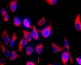
MitoOrange™ behaves like the potentiometric/classic lipophilic mitochondrial probes you already know: it preferentially accumulates in polarized mitochondria, giving you a bright, contiguous network (tubules, branches, and sometimes perinuclear “cloud”) that tracks organelle mass and membrane integrity. Because it’s orange-shifted, it also plays nicely with GFP/EGFP/FITC filters—you can keep your green channel free for a reporter or viability probe while mitochondria sit in the TRITC/Orange cube.
Hoechst 33342 binds AT-rich minor grooves of DNA in live cells (it’s permeant and widely used for live nuclear masking), lighting up nucleoplasm with that crisp blue signature under DAPI/UV excitation. The combination—Blue nucleus + Orange mitochondrion—is a workhorse look for a reason: high contrast, fast interpretation, and easy segmentation in analysis software.
A Protocol So Short You’ll Forget It Used to Be Annoying
Abbkine documents the practical workflow clearly; in practice it looks like this (always fine-tune dilution/time to your cell type):
- Prepare staining solution: dilute MitoOrange™ (1000×) and optionally Hoechst 33342 into 1× Assay Buffer (or low-serum medium if your cells tolerate it) to your working concentration (kit-style “pre-experiment titration” is encouraged).
- Incubate live cells (usually ~15–45 min, 37 °C, protected from light).
- Wash gently with buffer/medium to remove excess dye.
- Image immediately, or: fix with formaldehyde (many protocols fix right after the live stain so MitoOrange™ signal is retained), then proceed to permeabilization/blocking/immunostaining if you want extra markers.
For suspension cells, the same logic applies—spin down, resuspend in stain, incubate, wash, and either read on a plate reader/flow cytometer or cytospin/image.
Where KTC4005 Earns Its Keep: Applications That Matter
- Drug-induced mitochondrial toxicity & stress models
Instead of guessing, you get a per-cell read: swollen/rounded mitochondria + nuclear condensation → early stress; fragmented networks + chromatin margination → later-stage injury. Pair with a plate-read to rank compounds by “mitochondrial disruption index.” - Apoptosis / necrosis / ferroptosis-style QC
Nuclear condensation/blebbing + loss of contiguous mitochondrial reticular signal is a fast visual classifier while you wait for the heavier assays (caspases, Annexin V, etc.). - Stem cell & differentiation pipelines
MitoOrange™ intensity and network topology often shift during lineage commitment; having a stable nuclear mask lets you normalize signal and quantify %-positive/area-per-cell properly. - Immunofluorescence co-labeling without sacrificing a channel
Live/Post-fix mitochondrial blueprint from MitoOrange™ + a secondary-only-clean antibody in green/far-red = three informative channels without spectral gymnastics. - Flow cytometry (yes, really)
Because both dyes are live-permeant and strong, you can use Blue (Hoechst) vs. Orange (MitoOrange) scatter/dot plots to gate singlets, exclude debris, and see population-level ΔΨm/organelle-mass shifts—handy for quick hit/no-hit calls before you deep-dive with antibodies.
Why Labs Stick With This Kit (Instead of Reinventing the Wheel Weekly)
• Consistency: 1000× stocks + defined buffer beat “pipette a little extra from a DMSO stock because today feels right.”
• Speed: 15–45 min stain, wash, image. No transfections. No antibody incubations.
• Dual-platform value: Same preparation informs both microscopy/HCS and, when configured right, flow/plate reads.
• Fixed-retention option: Enables “live stain → fix → expand the panel” instead of forcing you to choose between dynamics and co-labeling.
• Storage/Logistics: Stable ≥6 months under recommended storage from shipment; ice-pack (blue-ice) shipping lowers hassle for typical lab freezers.
One-line positioning you can borrow: KTC4005 doesn’t try to be flashy—it tries to be the least noisy, most repeatable, fastest way to put a nucleus and a mitochondrial network into the same frame so your biology (not your staining) becomes the story.
Product Reference: KTC4005 – TraKine™ Mitochondrion and Nuclear Staining Kit
Learn more / download the manual / order: https://www.abbkine.com/product/trakine-mitochondrion-and-nuclear-staining-kit-ktc4005/


