


Beyond the Green Channel: Orange Fluorescence Mitochondrial Staining for Cleaner, Multiplex-Ready Live-Cell Imaging
Every fluorescence microscope tells its best stories in color—but only if those colors don't step on each other's toes. In live-cell mitochondrial imaging, the community has long defaulted to green-emitting dyes (FITC/GFP-filter compatible) because they're familiar, easy, and "just work." The problem? Modern experiments are almost never single-color anymore. Between GFP-tagged proteins, FITC-conjugated secondary antibodies, calcein-AM viability assays, and nuclear stains competing for the blue/green window, the spectral real estate around 490–530 nm is overcrowded. That's precisely where the TraKine™ Mitochondrion Staining Kit (Orange Fluorescence, KTC4004) from Abbkine earns its place. Built around a cell-permeant, cationic orange-emitting mitochondrial probe, this kit gives you a bright, ΔΨm‑dependent mitochondrial signal that slots neatly into the TRITC/Orange/RFP filter set—freeing up the green channel for your reporter, your antibody panel, or your viability dye. The result is a cleaner, more honest multiplex image, with less bleed-through, less compromise, and zero fixation required.
Why "Orange" Isn't Just Cosmetic—It's a Spectral Strategy
Mitochondrial tracking dyes—especially the potentiometric, cationic class—depend on the organelle's highly negative inner membrane potential (ΔΨm, typically −140 to −180 mV) to drive accumulation inside the matrix. The classic green options (e.g., rhodamine-based derivatives adapted for live imaging) absolutely get the job done, but they also anchor you to a spectral neighborhood that's already saturated in most multi-color protocols. Shifting the emission to the orange window (~560–590 nm excitation / ~570–620 nm emission, depending on the specific fluorophore design) does three pragmatic things at once: First, it opens the 488 nm laser / FITC/GFP channel so you can run a GFP-tagged construct, a green viability probe, or a green immunostaining panel in the same cell without fighting bleed-through. Second, it improves visual contrast in many brightfield + fluorescence overlay views because orange stands out against cytoplasmic autogreen. Third, on many confocal and widefield systems, the orange/red detection path carries less cellular autofluorescence background than the blue/green window, giving you a higher effective signal-to-noise ratio for fine mitochondrial tubules and branch points.
The Chemistry Under the Hood: ΔΨm‑Driven, Live-Cell Friendly, and Retention-Stable
The probe in TraKine™ KTC4004 is a lipophilic, monovalent cationic fluorophore designed to cross the plasma membrane readily, accumulate in polarized mitochondria, and stay put long enough to image—without aggressively collapsing the potential you're trying to visualize. Like its relatives in the mitochondrial dye family, it distributes across the inner membrane according to the Nernst equation; the more negative the matrix, the more dye piles in. That means intensity isn't just a shape overlay—it's semiquantitative: healthy, polarized mitochondria glow brighter; depolarized mitochondria (FCCP/CCCP-treated controls) lose signal. What distinguishes a well-formulated kit like this one is the balance: the dye concentration is tuned so you get strong tubular labeling at low nanomolar effective ranges, without the overt cytotoxicity, swelling artifacts, or runaway depolarization that come from overdosing cells. The formulation is also chosen for photostability in live imaging, so your time-lapse of mitochondrial dynamics doesn't fade to gray by frame 20.
Four Practical Reasons Labs Switch (or Add) the Orange Option
- Multiplexing without the headache. You can run an orange mitochondrial outline alongside a GFP-tagged protein (e.g., Drp1-GFP, MFN2-GFP, or GFP-LC3 if you're checking mitophagy contexts), plus a blue Hoechst/DAPI nuclear counterstain, and keep each channel clean. No spectral unmixing wizardry required on most standard filter sets.
- Fixable workflows when you need them. While the kit is optimized for live-cell use, the orange signal from this class of dye is often retainable through gentle paraformaldehyde fixation, which means you can stain live → fix → then run immunostaining (e.g., anti-TOM20, anti-COX IV, or an antibody against your protein of interest) in the same sample. That's a huge efficiency win for co-localization studies.
- Suspension cells and adherent cells alike. Because it's a small-molecule stain rather than an antibody, you don't fight accessibility issues—it works equally well on loosely attached or suspension formats (PBMCs, isolated primary neurons, or cultured cardiomyocytes in suspension preps) as long as you respect wash steps.
- Microplate-reader friendly. Beyond microscopy, the orange emission can be read on fluorescence plate readers equipped with TRITC/Orange filter sets, letting you turn mitochondrial potential into a semi-high-throughput endpoint for compound toxicity or metabolic stress screens—something purely imaging-based setups can't do at scale.
A 20-Minute Workflow That Doesn't Fight You
You don't need a 10-step ritual or a specialized perfusion chamber to get publication-grade mitochondrial outlines. The practical sequence with KTC4004 looks like this:
• Dilute the provided dye stock into pre-warmed, serum-free medium or assay buffer at the recommended working concentration (optimize 25–100 nM range depending on cell type).
• Replace the cell's medium with the dye solution and return to 37 °C / 5% CO₂ for 5–15 min (most lines look great at 10 min; suspension cells may prefer a short spin/wash step).
• Wash 1–2× with warm buffer or complete medium to remove excess dye.
• Image immediately on a TRITC/orange filter set (Ex/Em ~549/575 nm class), or fix gently if you're heading into a secondary staining step.
Because the dye is live-cell compatible at recommended concentrations, your cells keep dividing, migrating, and behaving normally through the session—so you can push into time-lapse (mitochondrial fission/fusion dynamics) or live pharmacological perfusion (watch ΔΨm collapse with FCCP in real time) without wondering if the dye itself tanked the cell.
Where This Kit Pulls Its Weight: Applications that Matter
Application What the Orange Mitochondrial Stain Gives You
Mitochondrial morphology & dynamics Crisp tubule/fragmentation readouts in live cells—Drp1/Fis1 overexpression, OPA1 knockdown, starvation, or mitophagy models
ΔΨm integrity & cytotoxicity Side-by-side with FCCP/CCCP/oligomycin controls → intensity shift = depolarization signature
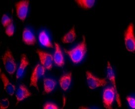
Multiplex live imaging Orange mitochondria + GFP reporter + blue nucleus = 3-channel story in one shot
Immuno-co-labeling (post-fix) Stain live → fix → antibody against TOM20/cytochrome c/your target → double-map structure vs. protein
Plate-reader screens Orange signal as a proxy for mitochondrial potential in compound libraries or toxicology batches
Cardiomyocyte & neuron prep QC Fast visual check of network integrity and mitochondrial health before you commit to downstream omics
The Bottom Line: Pick the Color That Fits Your Experiment, Not Just the Lab Tradition
There's a reason fluorescence microscopy moved from "one color at a time" to full-panel thinking—because biology is multivariate, and your staining should reflect that. The TraKine™ Mitochondrion Staining Kit (Orange Fluorescence, KTC4004) exists so you don't have to wedge every experiment into the green channel and then spend your afternoon subtracting bleed-through in ImageJ or FIJI. It gives you a bright, ΔΨm-sensitive, live-cell-ready mitochondrial signal that plays nicely with GFP reporters, blue nuclear stains, and far-red secondary panels—backed by Abbkine's formulation discipline so you're not debugging the dye instead of the science.
If your current mitochondrial imaging feels crowded, washed-out, or channel-limited, the fix might not be a new microscope—it might just be the right orange.
Product Reference: KTC4004 – TraKine™ Mitochondrion Staining Kit (Orange Fluorescence)
Learn more and order: https://www.abbkine.com/product/trakine-mitochondrion-staining-kit-orange-fluorescence-ktc4004/


