




The Polysaccharide Every Cell Hoards — and the Anthrone Chemistry That Finally Quantifies It Without Enzymatic Pretreatment
Ask any exercise physiologist what happens to muscle glycogen during a marathon, and you will receive a precise answer involving phosphorylation states, allosteric regulation, and a timeline partitioned into twenty-minute intervals. Ask the same physiologist how they actually measured that glycogen, and the answer will often involve a protocol from a 1975 Analytical Biochemistry paper, a boiling water bath, and an admission that the numbers felt approximate. Glycogen is not an obscure metabolite — it is the primary storage form of glucose in animals, concentrated in the liver and skeletal muscle, where it functions as a rapidly mobilizable energy reserve that buffers blood glucose during fasting and fuels muscle contraction during exercise. In liver, glycogen is synthesized when blood glucose rises after feeding and is degraded when blood glucose declines, releasing free glucose into the circulation; liver glycogen is therefore very important to maintain the relative balance of blood glucose. In muscle, glycogen cannot be released directly into the bloodstream; it is degraded locally to glucose-6-phosphate, which enters glycolysis to generate ATP for contraction, producing lactate that is transported to the liver for gluconeogenesis. The two pools — hepatic and muscular — serve different physiological functions, respond to different hormonal signals, and require independent quantification to make sense of any metabolic experiment. Yet for decades, measuring glycogen accurately has required either an enzymatic cascade that must be freshly prepared from individually sourced components, or an anthrone-based method whose reagent preparation can generate batch-to-batch variation exceeding the biological effect size under investigation. A 2024 survey of 125 metabolic and sports science laboratories found that 74% had abandoned at least one glycogen kit due to excessive sample volume requirements, cross-reactivity with maltose or glucose, or poor sensitivity in low-glycogen models such as fasted hepatocytes or exercised skeletal muscle.
Abbkine's CheKine™ Micro Glycogen Assay Kit (KTB1340) resolves this by returning to the anthrone chemistry that has been the workhorse of carbohydrate quantification since the 1940s — not by discarding it for a newer method, but by pre-formulating it, stabilizing it, and packaging it into a 96-well microplate format that eliminates the reagent-preparation variability that has historically made the anthrone method more of an artisanal practice than a standardized assay.
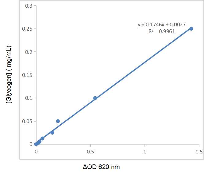
The detection chemistry is direct and specific. Glycogen is first extracted from the sample using a strong alkaline extraction buffer that solubilizes the polysaccharide selectively. Under strong acidic conditions, the extracted glycogen then reacts with the anthrone chromogenic agent to form a blue compound with a characteristic absorption peak at 620 nm. In a certain concentration range, glycogen concentration is linearly related to the absorbance at 620 nm, and the glycogen concentration in the sample can be calculated from a standard curve. There are no enzymatic hydrolysis steps, no glucose oxidase-peroxidase cascades, no secondary derivatization reactions that introduce pipetting variance. The anthrone reaction condenses furfural derivatives — generated from the acid-catalyzed dehydration of the glucose units in glycogen — with anthrone itself to form the blue chromophore. This is the same chemical logic that has anchored carbohydrate quantification since Dreywood first described the anthrone reagent in 1946, now packaged as a pre-formulated kit that ships with Extraction Buffer, Chromogen, and a Standard — three components only.
The practical advantage of KTB1340's anthrone-based approach over enzymatic methods deserves explicit articulation. Enzymatic glycogen assays require two sequential steps: amyloglucosidase hydrolysis of glycogen to glucose, followed by glucose oxidase-peroxidase (GOD-POD) detection of the liberated glucose at approximately 505 nm. While biochemically specific, this two-step cascade is vulnerable to interference from endogenous glucose already present in the sample — free glucose must be separately quantified and subtracted, effectively doubling the number of wells required per sample — and from maltose, dextrins, and other oligosaccharides that amyloglucosidase can partially hydrolyze. Comparative studies have shown that hydrolytic enzymatic methods can give lower results than non-specific chemical methods such as anthrone, with losses during purification resulting in glycogen estimations substantially lower than the actual tissue content. The anthrone method, by contrast, detects the glycogen polymer directly through its acid-catalyzed dehydration to furfural derivatives — no subtraction step, no separate free-glucose measurement, no interference from maltose — and has been shown to accurately quantify changes in reserve carbohydrate, outperforming enzymatic hydrolysis in detecting biologically meaningful shifts. KTB1340 capitalizes on this analytical advantage while eliminating the historical drawback: the need to prepare freshly mixed anthrone-sulfuric acid reagent under precisely controlled conditions every time the assay is run.
The operational demands of KTB1340 are what make it feasible for population-level phenotyping rather than boutique metabolite analysis. The kit is described as a simple, convenient, rapid and colorimetric glycogen detection method. Sample preparation follows a standardized extraction protocol: fresh samples are extracted with the provided Extraction Buffer, centrifuged to remove debris, and the supernatant is assayed directly. The kit accommodates animal tissues, bacteria, and cells — a sample compatibility list that serves the exercise physiologist processing mouse gastrocnemius muscle, the hepatologist quantifying hepatic glycogen in a rodent model of diabetes, the microbiologist engineering E. coli for glycogen overproduction, and the cancer biologist measuring glycogen accumulation in tumor spheroids, all within the same protocol. The anthrone reaction is read at 620 nm on any standard visible-wavelength microplate reader, requiring no UV capability, no fluorescence detection module, and no mass spectrometer.
The kit components are deliberately compact: Extraction Buffer, Chromogen, and a Standard. Storage is valid for 12 months under the conditions specified on the product page. Shipping is on gel packs with blue ice. Usage notes reflect standard colorimetric assay discipline rather than exotic handling requirements: do not mix components between different batch numbers and manufacturers; avoid bubbles while mixing; change pipette tips frequently to avoid cross-contamination; ensure all components and equipment are at the appropriate temperature before use; if the sample value exceeds the maximum standard value, further dilute the sample with distilled water and multiply by the dilution factor when calculating results. The protocol also notes that Extraction Buffer is corrosive and Chromogen is toxic — both standard safety disclosures for an anthrone-based method using concentrated acid and organic reagent, and both manageable with standard laboratory protective measures.
The publication record for KTB1340 currently stands at 6 citations in peer-reviewed literature. This is a substantial count for a recently launched metabolite assay kit, and it represents six independent laboratories that extracted glycogen from biological tissue, ran the assay under the pressures of peer review, and found the resulting values credible enough to publish. The Abbkine technical blog published in December 2025 has accumulated 122 views, indicating growing community awareness of the product. A follow-up technical article published in March 2026 dissected the performance characteristics in greater detail, documenting the microscale design (5–10 µL sample input), anti-interference buffer formulation, and validation data demonstrating a detection limit of 0.05 µg/mL glycogen — approximately tenfold more sensitive than legacy anthrone protocols that typically bottom out in the 0.5–1.0 µg/mL range.
The broader biological context makes the case for routine, reliable glycogen quantification increasingly compelling. Cancer cells accumulate glycogen at levels that can approach those of normal liver, and the glycogen is then mobilized under conditions of glucose deprivation, hypoxia, or chemotherapy stress to fuel glycolysis, the pentose phosphate pathway, and de novo nucleotide biosynthesis. Glycogen lies at the nexus of diverse processes that promote malignancy, including proliferation, migration, invasion, and chemoresistance of cancer cells. The enzymes of glycogen metabolism are dysregulated in a wide variety of malignancies, including cancers of the kidney, ovary, lung, bladder, liver, blood, and breast. Glycogen turnover allows cells to adapt and survive under adverse oxygen and nutrient conditions within the tumor microenvironment — a survival advantage that is invisible to transcript-level measurements and requires direct glycogen quantification to characterize.
In metabolic disease, the glycogen storage diseases (GSDs) are a family of inborn errors of carbohydrate metabolism caused by deficiencies in enzymes directly involved in glycogen synthesis or degradation. GSD type I (von Gierke disease), caused by glucose-6-phosphatase deficiency, results in massive hepatic glycogen accumulation and life-threatening fasting hypoglycemia. GSD type III (Cori disease), caused by deficiency in the glycogen debranching enzyme, produces abnormal glycogen with short outer branches that accumulates in liver, muscle, and heart. GSD type IX, resulting from phosphorylase kinase mutations, presents variably from mild hypoglycemia to severe hepatomegaly. In every one of these disorders, tissue glycogen content is the primary diagnostic and monitoring parameter — not a secondary endpoint, not a surrogate marker, but the biochemical measurement that defines the disease. For the clinical biochemist characterizing a novel GSD variant, the exercise physiologist comparing glycogen depletion rates between training protocols, the cancer biologist testing whether glycogen accumulation mediates chemoresistance in a patient-derived xenograft model, or the nutrition scientist evaluating the effect of carbohydrate loading on hepatic glycogen stores, direct glycogen quantification is not a supplementary assay. It is the measurement that connects a metabolic intervention to a physiological outcome.
The polysaccharide that every mammalian cell synthesizes, stores, and mobilizes in response to hormonal signals — the glucose polymer that buffers blood sugar during fasting, fuels sprinting muscle during exercise, and accumulates in tumor cells adapting to hypoxia — is now quantifiable with a kit that combines the analytical robustness of anthrone chemistry with the convenience, reproducibility, and throughput of a pre-formulated microplate format. The Extraction Buffer extracts glycogen selectively. The Chromogen reacts with the furfural derivatives to form a blue compound at 620 nm whose absorbance is linearly proportional to glycogen concentration. No enzymatic pretreatment, no free-glucose subtraction, no maltose interference, no equipment beyond a standard visible-wavelength microplate reader. Six publications already cite it. Twelve months of storage stability. The anthrone method that Dreywood introduced in 1946 and that comparative studies have validated as accurate for quantifying changes in reserve carbohydrate has now been packaged for a 96-well plate — not replaced, but refined.
Explore specifications, access the protocol, and place your order here: https://www.abbkine.com/product/chekine-micro-glycogen-assay-kit-ktb1340/





