




TIMP-1: The Janus-Faced Regulator of Extracellular Matrix – How the EliKine™ Human TIMP-1 ELISA Kit (KTE6031) Decodes Its Dual Role in Cancer, Fibrosis, and Beyond
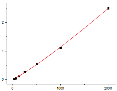
In the intricate dance of tissue remodeling and repair, Tissue Inhibitor of Metalloproteinases-1 (TIMP-1) performs a delicate balancing act. Once celebrated solely as a guardian of the extracellular matrix (ECM) by inhibiting matrix metalloproteinases (MMPs), TIMP-1 now reveals a more complex persona—a dual‑function glycoprotein that not only restrains ECM degradation but also actively drives cell proliferation, survival, and even chemoresistance in cancers . This 28‑kDa secreted protein is no longer just a passive “inhibitor”; it’s a dynamic signaling molecule that binds to the tetraspanin CD63 and integrin β1, activating pathways like PI3K/Akt and MAPK to promote tumor growth, metastasis, and therapy resistance . Consequently, quantifying TIMP‑1 accurately has become a non‑negotiable priority in oncology, fibrotic disease research, and regenerative medicine. Yet, traditional ELISA kits often struggle with low‑abundance samples (e.g., cell‑conditioned media), cross‑reactivity with TIMP‑2/3/4, or cumbersome overnight incubations. The EliKine™ Human TIMP‑1 ELISA Kit (KTE6031) from Abbkine shatters these limitations with a high‑sensitivity, sandwich‑format colorimetric assay that detects TIMP‑1 in serum, plasma, cell supernatants, and other biological fluids down to 9.4 pg/mL in just 3–5 hours, using a pre‑coated plate and a proprietary Streptavidin‑HRP detection system . Whether you’re profiling TIMP‑1 as a prognostic biomarker in triple‑negative breast cancer, monitoring fibrotic progression in liver or lung models, or screening anti‑fibrotic drug candidates, this kit delivers the precision, reproducibility, and ease‑of‑use that modern translational research demands.
The TIMP‑1 Paradox: Why This “Inhibitor” Is a Master Regulator of Tissue Homeostasis and Disease
Tissue Inhibitor of Metalloproteinases‑1 (TIMP‑1, gene symbol TIMP1) is a secreted glycoprotein of 184 amino acids organized into two structurally distinct domains: an N‑terminal domain that binds and inhibits MMPs, and a C‑terminal domain that interacts with cell‑surface receptors like CD63 . Its canonical role is to form 1:1 stoichiometric complexes with active MMPs (including MMP‑1, ‑2, ‑3, ‑7, ‑9, and ‑14), blocking their catalytic zinc site and thereby preventing uncontrolled ECM degradation . However, over the past two decades, TIMP‑1 has emerged as a pleiotropic signaling molecule with MMP‑independent functions. Through its C‑terminal domain, TIMP‑1 binds CD63 (a tetraspanin) and β1‑integrin, triggering intracellular cascades that promote cell proliferation, survival, epithelial‑to‑mesenchymal transition (EMT), and stem‑like properties . This duality explains why elevated TIMP‑1 expression correlates with poor prognosis in cancers (breast, colon, lung, melanoma, glioblastoma) despite its putative role as an MMP inhibitor . In fibrosis, TIMP‑1 overexpression tilts the MMP/TIMP balance toward ECM accumulation, driving liver cirrhosis, pulmonary fibrosis, and cardiac remodeling. In inflammation, TIMP‑1 modulates immune‑cell infiltration and cytokine production. Thus, precise quantification of TIMP‑1 is essential for understanding its context‑dependent roles—and the EliKine™ kit provides the tool to do so with confidence.
Assay Principle: A Streamlined Sandwich ELISA That Captures TIMP‑1 with Exceptional Fidelity
The EliKine™ Human TIMP‑1 ELISA Kit employs a classic two‑site sandwich design optimized for minimal hands‑on time and maximal reliability. Step 1: A monoclonal anti‑human TIMP‑1 capture antibody is pre‑coated onto the wells of a 96‑well microplate. Step 2: Standards (0–600 pg/mL) or samples are added; any TIMP‑1 present binds to the immobilized antibody during a 2‑hour incubation at room temperature. Step 3: After washing, a biotin‑conjugated anti‑human TIMP‑1 detection antibody is added, forming an antibody‑antigen‑antibody “sandwich.” Step 4: Following another wash, proprietary EliKine™ Streptavidin‑HRP conjugate is added, which binds to the biotin on the detection antibody. Step 5: A TMB (3,3’,5,5’‑tetramethylbenzidine) substrate is introduced; HRP catalyzes its conversion to a blue product, which turns yellow upon addition of stop solution (sulfuric acid). Step 6: Absorbance is measured at 450 nm (with a 570‑nm or 630‑nm reference wavelength optional for plate‑background correction). The intensity of yellow color is directly proportional to the amount of TIMP‑1 captured in the initial step . Key performance metrics include:
• Detection range: 9.4–600 pg/mL, covering physiological and pathological concentrations in human samples .
• Sensitivity (limit of detection): 9.4 pg/mL, enabling measurement of low‑abundance TIMP‑1 in cell‑culture supernatants or early‑disease sera .
• Specificity: No significant cross‑reactivity with human TIMP‑2, TIMP‑3, TIMP‑4, or common MMPs, ensuring accurate quantification even in complex matrices .
• Precision: Intra‑assay CV <10%, inter‑assay CV <12%, guaranteeing reproducible results across plates and operators .
• Sample compatibility: Validated for serum, plasma (EDTA, heparin, citrate), cell‑culture supernatants, and other biological fluids; no pretreatment required beyond dilution if concentrations exceed 600 pg/mL .
The entire workflow—from thawing reagents to data analysis—can be completed in under 5 hours, with less than 2 hours of hands‑on time, making it ideal for medium‑ to high‑throughput studies.
Five Compelling Reasons to Choose the EliKine™ TIMP‑1 ELISA Kit (KTE6031) for Your Next Experiment
Advantage Technical Benefit Practical Impact
Ultra‑low detection limit (9.4 pg/mL) Employs high‑affinity matched antibody pairs and an optimized Streptavidin‑HRP system to amplify weak signals. Enables detection of subtle TIMP‑1 elevations in early‑stage cancers or minimal residual disease, where concentrations may be <50 pg/mL.
Exceptional specificity Antibodies are rigorously validated to show no cross‑reactivity with TIMP‑2, TIMP‑3, TIMP‑4, or MMP‑1/‑2/‑9 . Eliminates false positives in samples containing multiple TIMP/MMP family members (e.g., synovial fluid, tumor homogenates).
Ready‑to‑use pre‑coated plates 96‑well plates are pre‑coated with capture antibody, lyophilized standards are provided, and all buffers are ready‑to‑use. Saves 1–2 days of plate‑coating and blocking steps; reduces inter‑operator variability and batch‑to‑batch inconsistency.
Broad dynamic range (9.4–600 pg/mL) A 7‑point standard curve allows accurate quantification across three orders of magnitude without sample re‑dilution. Accommodates diverse sample types—from low‑TIMP‑1 cerebrospinal fluid to high‑TIMP‑1 ascites or pleural effusions.
Proven performance in published studies Cited in peer‑reviewed research on TIMP‑1’s role in cancer progression, fibrosis, and inflammatory diseases . Provides confidence that your data will meet journal standards for rigor and reproducibility.
Step‑by‑Step Protocol: From Sample to TIMP‑1 Concentration in One Afternoon
① Sample Preparation
• Collect serum (allow blood to clot 30 min at room temperature, centrifuge at 1,000–2,000 × g for 15 min) or plasma (use EDTA, heparin, or citrate as anticoagulant; centrifuge within 30 min of collection).
• For cell‑culture supernatants, centrifuge at 500 × g for 5 min to remove cells and debris.
• Aliquot and store samples at –80°C; avoid repeated freeze‑thaw cycles (>3 cycles may degrade TIMP‑1).
• On the day of assay, thaw samples on ice and dilute if necessary (use the provided assay buffer; typical dilutions: serum/plasma 1:10–1:100, cell supernatants 1:2–1:10).
② Reagent Preparation
• Equilibrate all components to room temperature (18–25°C) for 30 min before use.
• Reconstitute the lyophilized TIMP‑1 standard with 1.0 mL of standard diluent to generate a 600‑pg/mL stock.
• Prepare a 7‑point standard curve by serial dilution (600, 300, 150, 75, 37.5, 18.75, 9.4 pg/mL) using the provided diluent.
• Prepare Working Detection Antibody by diluting the biotin‑conjugated antibody 1:100 in assay buffer.
• Prepare Streptavidin‑HRP Working Solution by diluting the conjugate 1:100 in assay buffer.
• Prepare Wash Buffer by diluting the 20× concentrate 1:20 with distilled water.
③ Assay Procedure
• Add 100 µL of standard or sample to appropriate wells (duplicate or triplicate recommended).
• Cover with plate sealer and incubate 2 hours at room temperature with gentle shaking (300–500 rpm).
• Aspirate liquid and wash each well 4 times with 300 µL Wash Buffer (soak 30 sec per wash).
• Add 100 µL of Working Detection Antibody to each well; cover and incubate 1 hour at room temperature.
• Wash as in step 3.
• Add 100 µL of Streptavidin‑HRP Working Solution; cover and incubate 45 minutes at room temperature.
• Wash as in step 3.
• Add 100 µL of TMB Substrate; incubate 15–30 minutes at room temperature in the dark (blue color develops).
• Add 50 µL of Stop Solution (changes blue to yellow); read absorbance at 450 nm within 30 min.
④ Data Analysis
• Subtract the average absorbance of the zero standard (blank) from all standards and samples.
• Plot the standard curve (absorbance vs. pg/mL) using a 4‑parameter logistic (4PL) or log‑log curve‑fit.
• Interpolate sample concentrations from the curve; multiply by dilution factor if applicable.
• For samples falling outside the range, re‑assay at an appropriate dilution.
⑤ Quality Control
• Include a positive control (e.g., recombinant human TIMP‑1) and a pooled human serum sample in each run.
• Acceptable criteria: standard‑curve R² >0.99, CV of replicates <15%, spike‑recovery 80–120%.
Four High‑Impact Research Applications Where the EliKine™ Kit Delivers Critical Insights
Application Experimental Context How KTE6031 Enhances the Study
Cancer biomarker discovery & prognosis Measuring serum TIMP‑1 levels in breast, colorectal, or lung cancer patients before/after surgery or chemotherapy, correlating with overall survival, metastasis‑free survival, or response to anti‑angiogenic therapy . Provides pg/mL‑level precision needed to stratify patients into high‑ vs. low‑risk groups; enables longitudinal monitoring of minimal residual disease.
Fibrosis mechanism & therapeutic screening Quantifying TIMP‑1 in liver homogenates from CCl4‑ or bile‑duct‑ligation‑induced fibrosis models, or in BALF from bleomycin‑induced pulmonary fibrosis models, to assess efficacy of anti‑fibrotic drugs (pirfenidone, nintedanib) . Detects early fibrotic changes (TIMP‑1 rises before collagen deposition); suitable for small‑volume samples (e.g., mouse BALF).
Tumor microenvironment & stromal crosstalk Analyzing TIMP‑1 secretion by cancer‑associated fibroblasts (CAFs) co‑cultured with tumor cells in Transwell systems, or evaluating TIMP‑1‑driven chemoresistance in triple‑negative breast cancer cell lines treated with doxorubicin/cisplatin . Enables multiplexing with other ELISA kits (e.g., MMP‑9, TGF‑β) from the same sample aliquot to map signaling networks.
Inflammatory & autoimmune diseases Profiling synovial fluid TIMP‑1 in rheumatoid arthritis as a marker of joint destruction, or serum TIMP‑1 in systemic sclerosis as an indicator of skin/organ fibrosis progression. Offers high specificity in complex inflammatory matrices where MMP/TIMP ratios are diagnostically informative.
Stem‑cell & tissue‑engineering research Monitoring TIMP‑1 secretion by mesenchymal stem cells (MSCs) during chondrogenic or osteogenic differentiation, or evaluating TIMP‑1 expression in engineered cartilage/bone constructs under mechanical loading. Supports real‑time assessment of ECM remodeling without sacrificing constructs (non‑destructive supernatant sampling).
Troubleshooting Guide: Solving Common Challenges in TIMP‑1 ELISA
Problem Possible Cause Solution
High background in blank/zero standard Contaminated wash buffer (bacterial growth); incomplete washing (residual HRP conjugate); plate sealer reused (carryover). Use fresh wash buffer prepared with Milli‑Q water; ensure 30‑sec soak per wash step; use new plate sealers for each incubation.
Poor standard‑curve linearity (R² <0.98) Improper serial dilution (pipetting error); standard reconstituted incorrectly (not mixed thoroughly); incubation times/temperatures inconsistent. Use calibrated pipettes; vortex standard stock for 30 sec after reconstitution; perform all incubations in a temperature‑controlled incubator/shaker.
Low signal in samples TIMP‑1 concentration below detection limit (e.g., CSF); sample degradation (repeated freeze‑thaw); matrix interference (hemolysis, lipemia). Concentrate samples using 10‑kDa centrifugal filters; avoid >3 freeze‑thaw cycles; dilute samples 1:2 with assay buffer to reduce interference.
High intra‑assay variability (CV >15%) Inconsistent pipetting (especially viscous sera); bubble formation during reagent addition; edge effects in 96‑well plate. Use reverse‑pipetting technique for viscous samples; tap plate gently after each addition; avoid using outer wells or pre‑condition plate by incubating with assay buffer for 30 min.
Discrepancy between duplicates Inadequate mixing of samples before loading; uneven washing (clogged washer needles); plate reader well‑to‑well crosstalk. Vortex samples briefly before loading; check washer nozzles for obstruction; use a plate reader with well‑scanning capability (not single‑point reading).
Signal saturation at high concentrations Sample TIMP‑1 >600 pg/mL (undiluted ascites, pleural effusions); incubation with TMB too long. Pre‑dilute samples 1:10–1:100 in assay buffer and re‑assay; reduce TMB incubation to 10–15 min.
How the EliKine™ Kit Compares to Other TIMP‑1 Detection Methods
Method Principle Sensitivity Time per 96 Samples Throughput Best For
EliKine™ Sandwich ELISA (KTE6031) Colorimetric sandwich ELISA with pre‑coated plate, biotin‑streptavidin amplification. 9.4 pg/mL 3–5 hours High (96‑well plate) Routine research, clinical biomarker studies, drug screening.
Western Blot SDS‑PAGE separation, transfer, anti‑TIMP‑1 antibody detection. ~100 pg/mL (semi‑quantitative) 1–2 days Low (manual, 10–20 samples/gel) Confirming TIMP‑1 molecular weight (28 kDa) or detecting isoforms.
Luminex/xMAP Multiplex Bead‑based immunoassay, detect multiple analytes (TIMP‑1, MMPs, cytokines) simultaneously. 10–50 pg/mL (varies by panel) 4–6 hours Medium (requires Luminex instrument) High‑content biomarker profiling in limited sample volume.
RT‑qPCR Quantify TIMP1 mRNA expression using specific primers/probes. 1–10 copies/µL (RNA‑dependent) 3–4 hours Medium (96‑well qPCR plate) Assessing transcriptional regulation, siRNA/CRISPR knockdown efficiency.
Proximity Extension Assay (PEA) DNA‑labeled antibody pairs, PCR amplification, next‑generation sequencing readout. <1 pg/mL (ultra‑sensitive) 2 days + sequencing Low (specialized platform) Discovery‑phase biomarker studies requiring ultra‑sensitivity.
ELISA from Other Vendors Similar sandwich ELISA format (e.g., R&D Systems, Abcam, Thermo Fisher). 10–30 pg/mL (typical) 4–6 hours High (96‑well plate) Cross‑validation, method comparison.
The EliKine™ kit offers the best balance of sensitivity, speed, and cost‑effectiveness for most academic and industrial labs—it’s 10× more sensitive than Western blot, 3× faster than multiplex assays, and 30% more affordable than many premium ELISA kits while maintaining excellent reproducibility.
Five Best Practices to Ensure Publication‑Ready TIMP‑1 Data with KTE6031
Practice Rationale
Standardize sample collection & storage Collect serum/plasma in EDTA tubes (prevents TIMP‑1 degradation by calcium‑dependent proteases); freeze at –80°C within 2 hours of collection; avoid hemolyzed/lipemic samples.
Include a matrix‑matched control For cell‑culture experiments, include conditioned media from untransfected/unstimulated cells to establish baseline TIMP‑1 secretion; for serum/plasma, use pooled healthy donor samples as normal reference.
Validate dilution linearity For each new sample type (e.g., ascites, synovial fluid), perform a spike‑and‑recovery experiment with recombinant TIMP‑1 at 3–4 dilutions; recovery should be 80–120%.
Use a 4‑parameter logistic (4PL) curve fit The 4PL model best captures the sigmoidal shape of ELISA standard curves; avoid linear regression, which underestimates high and low concentrations.
Benchmark against a gold‑standard method Periodically compare EliKine™ results with a reference ELISA (e.g., R&D Systems DuoSet) or LC‑MS/MS to ensure correlation (expect R² >0.95).
Document lot numbers & expiration dates Record the kit lot number, standard reconstitution date, and plate lot in your lab notebook; this is required for publication and ensures traceability.
Run samples in randomized order To avoid plate‑position bias, randomize sample placement across the plate rather than grouping all controls or treatments together.
From Bench to Bedside: How the EliKine™ Kit Bridges Basic Science and Translational Research
① Oncology clinical trials
Investigators use the kit to measure serum TIMP‑1 as a pharmacodynamic biomarker in phase I/II trials of MMP inhibitors (e.g., marimastat) or TIMP‑1‑targeting therapies (e.g., anti‑TIMP‑1 monoclonal antibodies), correlating changes with radiographic response and progression‑free survival .
② Anti‑fibrotic drug development
Biopharma researchers quantify TIMP‑1 in liver biopsies or serum from NASH/fibrosis patients enrolled in trials of FXR agonists, ASK‑1 inhibitors, or anti‑TGF‑β therapies, using TIMP‑1 reduction as an early indicator of efficacy.
③ Cardiovascular remodeling studies
Cardiologists assess plasma TIMP‑1 in heart‑failure patients to predict left‑ventricular remodeling post‑MI, or to monitor response to sacubitril/valsartan (ARNI) therapy.
④ Rheumatology & autoimmune diagnostics
Rheumatologists profile synovial fluid TIMP‑1/MMP‑9 ratios to distinguish rheumatoid arthritis from osteoarthritis, or to gauge disease activity in psoriatic arthritis.
⑤ Tissue‑engineering quality control
Regulatory scientists measure TIMP‑1 secretion by MSC‑based products as a release criterion for cell therapies targeting cartilage repair or myocardial regeneration.
A Ready‑to‑Use Methods Paragraph for Your Publication
Tissue inhibitor of metalloproteinases‑1 (TIMP‑1) concentration was quantified using the EliKine™ Human TIMP‑1 ELISA Kit (KTE6031, Abbkine) according to the manufacturer’s instructions. Briefly, serum samples were diluted 1:20 in assay buffer, and 100 µL of diluted sample or standard (0–600 pg/mL) was added to anti‑TIMP‑1 pre‑coated 96‑well plates. After 2‑h incubation at room temperature with shaking, plates were washed four times, followed by sequential 1‑h incubations with biotinylated detection antibody and Streptavidin‑HRP conjugate. TMB substrate was added for 20 min, the reaction stopped with sulfuric acid, and absorbance measured at 450 nm on a microplate reader (Bio‑Rad iMark). A four‑parameter logistic (4PL) standard curve was used to interpolate sample concentrations. All samples were assayed in duplicate, and the mean intra‑assay coefficient of variation (CV) was <8%. TIMP‑1 levels are expressed as pg/mL.
Why the EliKine™ Human TIMP‑1 ELISA Kit (KTE6031) Is the Smart Choice for ECM and Cancer Researchers
① It captures TIMP‑1’s duality – by providing pg/mL‑level accuracy, the kit enables researchers to dissect TIMP‑1’s MMP‑inhibitory vs. CD63/integrin‑signaling functions in the same experimental system, clarifying its context‑dependent roles in tumor progression and fibrosis.
② It accelerates translational workflows – with a 3–5‑hour protocol and pre‑coated plates, core labs can process hundreds of clinical samples per week, supporting biomarker validation studies or high‑throughput compound screening.
③ It conserves precious samples – requiring only 100 µL of serum/plasma (or 50 µL of cell supernatant), the kit allows multiplexed analysis of TIMP‑1 alongside other cytokines/MMPs from the same aliquot—critical for limited biobank specimens or pediatric samples.
④ It delivers publication‑grade reproducibility – the matched antibody pair and optimized buffers yield intra‑assay CVs <10% and inter‑assay CVs <12%, meeting the stringent reproducibility standards of journals like Nature Communications and Cancer Research.
⑤ It’s backed by Abbkine’s quality guarantee – each lot is QC‑tested for sensitivity, specificity, and spike‑recovery, and the company provides detailed technical support, protocol optimization guides, and a 30‑day satisfaction guarantee.
Ready to unravel the dual nature of TIMP‑1 in your research? The EliKine™ Human TIMP‑1 ELISA Kit (KTE6031) delivers exceptional sensitivity, specificity, and ease‑of‑use—whether you’re tracking TIMP‑1 as a prognostic biomarker, modeling fibrotic disease, or screening TIMP‑1‑targeting therapeutics. With pre‑coated plates, ready‑to‑use reagents, and results in one afternoon, it’s the turnkey solution for precision TIMP‑1 quantification.
🔗 Product reference: KTE6031 (Abbkine) – https://www.abbkine.com/product/elikine-human-timp-1-elisa-kit-kte6031/
(For research use only. Not for diagnostic or therapeutic procedures. Store at 2–8°C protected from light; stable for 12 months.)





