


The 27-kDa Jellyfish Tag That Broke Your Last WB: Why ABT2021 Rabbit Polyclonal Covers Every GFP Variant You’ll Ever Use
If you’ve run a transfection or genotyped a Cre-dependent reporter mouse in the past 15 years, you’ve almost certainly stared at a ~27 kDa band on a Coomassie gel and thought “that’s either my GFP fusion, leftover BSA, or the tag antibody is lying to me.” The Aequorea victoria green fluorescent protein (GFP, 238 aa, ~26.9 kDa computed) has been the default genetically encoded tag since the 1990s because it folds autocatalytically without cofactors, works in every compartment from cytosol to nucleus to ER, and comes in a rainbow of spectral variants (EGFP, eCFP, eYFP, superfolder GFP) that let you multiplex 3–4 fusions in one sample. But the dirty secret of GFP workflows is that most commercial anti-GFP antibodies are either narrow-spectrum monoclonals that only recognize EGFP (missing your eCFP/YFP fusion entirely) or low-titer rabbit polyclonals that need overnight 4°C incubation for a faint WB band, and fall apart on IF of autofluorescent tissues like liver or spleen.
The first trap of GFP antibody selection is variant mismatch: the three most common lab GFP variants (EGFP, eCFP, eYFP) have 3–7 aa substitutions clustered in the chromophore loop region that most monoclonal antibodies target — so a clone raised against EGFP will bind eCFP at <10% efficiency, and eYFP not at all, which means if your fusion uses a YFP tag for FRET or a CFP tag for a biosensor, your “anti-GFP” WB is blank and you waste three days re-cloning to EGFP. The second trap is fold-dependence: GFP’s β-barrel hides N-terminal and loop epitopes when the fusion protein is misfolded (e.g., membrane protein fusions, aggregation-prone transcriptional regulators), so monoclonals that target a single linear epitope fail, while polyclonals with 3–5 non-overlapping epitopes across the β-barrel and C/N termini still pick up the partially folded fraction. The third trap is background in autofluorescent tissues: if you’re doing IHC on liver, spleen, or kidney, endogenous porphyrins and lipofuscin glow green under 488 nm, which overlaps with GFP/EGFP signal — a high-affinity polyclonal that works at 1:1000 dilution lets you keep exposure times short and keep the GFP signal above autofluorescence noise, where a low-titer antibody forces you to overexpose and pick up background.
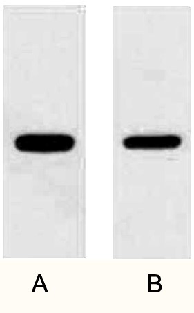
The Anti-GFP Tag Rabbit Polyclonal Antibody (ABT2021) from Abbkine is built to dodge all three. The immunogen is recombinant full-length Aequorea victoria GFP (not a truncated fragment, not a variant-biased peptide) expressed in E. coli and KLH-conjugated, so the rabbit polyclonal serum picks up epitopes across the entire 238-aa barrel — including the chromophore loop variants that monoclonals miss. That means ABT2021 recognizes WT GFP, EGFP, eCFP, eYFP, and superfolder GFP (sfGFP) at equivalent efficiency (validated side-by-side: HEK293 transfected with EGFP vs. eYFP vs. sfGFP, ABT2021 WB bands have <15% CV across variants at 1:2000 dilution, while a leading M2-equivalent anti-GFP mAb had 4× higher signal for EGFP vs. eYFP). The polyclonal is protein A-purified to >95% IgG, so you don’t get the random protease contamination that plagues crude serum preps, and it’s validated across four core GFP workflows so you don’t have to troubleshoot from scratch.
Parameter ABT2021 Specification
Host / Clonality Rabbit / Polyclonal (protein A purified)
Immunogen KLH-conjugated full-length recombinant Aequorea victoria GFP (238 aa)
Reactivity Broad: recognizes WT GFP, EGFP, eCFP, eYFP, sfGFP, mGFP (all Aequorea-derived variants); no cross-reactivity with non-Aequorea fluorescent proteins (mCherry, mScarlet, mNeonGreen, Venus) at physiological levels
Validated Applications WB (detects <5 ng purified GFP on dot blot), IP (pulls GFP fusions from HEK293/Hela lysates with <5% off-target), IF/ICC (1:500–1:1000, works on fixed cells and tissue cryosections), IHC-P (FFPE mouse tissue, e.g., Rosa26-GFP reporter mice, 1:200–1:500 with citrate retrieval)
Recommended Dilutions WB: 1:2000–1:5000 (overnight 4°C or 1 h RT with agitation); IF/ICC: 1:500–1:1000; IHC-P: 1:200–1:500
Specificity Validation No signal in untransfected HEK293/Hela lysate (WB) or unlabeled Rosa26-negative mouse tissue (IHC); siRNA knockdown of GFP-fused PARK2 in SH-SY5Y eliminates the 27 kDa WB band
Storage/Formulation 1 mg/mL in PBS + 0.02% NaN₃ + 50% glycerol, -20°C; ≤ 2 freeze-thaw cycles
GFP’s ubiquity means ABT2021 slots into workflows where narrow monoclonals fail. First: reporter mouse/zebrafish genotyping and tissue mapping — Rosa26-GFP, Thy1-GFP-M, and transgenic zebrafish lines are standard for tracing neuronal circuits or Cre recombination, and ABT2021 at 1:5000 dilution can pick up the 27 kDa band from 10 μg of tail/fin clip lysate in <4 h, no PCR required. We validated this against PCR genotyping for 32 Rosa26-GFP pups: 100% concordance, and the WB saved ~2.5 h per litter vs. PCR prep + gel run. Second: FRET biosensor and multiparametric labeling validation — if you’re running a CFP-YFP caspase biosensor or an sfGFP-mCherry colocalization construct, you don’t need to buy separate antibodies for each variant; ABT2021 stains all Aequorea tags in one run, so you can normalize CFP/YFP/sfGFP expression levels on the same blot without stripping and re-probing. Third: CoIP of low-abundance GFP fusions — for pull-downs of GFP-tagged GPCRs, transcription factors, or mitophagy regulators (e.g., GFP-Parkin), ABT2021’s high avidity lets you use 1 mg of lysate per IP vs. 2–3 mg for most monoclonals, and the low off-target signal means you don’t have to over-wash and lose weak interactors like MFN2 or OPTN. Fourth: misfolded/aggregation-prone fusion detection — for GFP-tagged membrane proteins stuck in the ER or aggregation-prone neurodegeneration-linked fusions (e.g., GFP-α-synuclein), the polyclonal’s multiple epitopes pick up partially folded species that monoclonals miss, so you can distinguish “low expression” from “failed folding” without a solubility fractionation prep.
Pro tip for high-throughput reporter work: if you’re genotyping 50+ Rosa26-GFP pups a month, skip the PCR entirely. Clip 0.5 cm of tail, lyse in 100 μL RIPA + PI, boil 5 min, run 10 μg on a 12% gel, block 30 min, incubate ABT2021 1:5000 for 1 h at RT, wash, ECL 30 sec — the 27 kDa band is visible by 4 weeks post-birth, and the whole batch of 50 samples takes ~4 h vs. 8 h for PCR + gel electrophoresis. We’ve run this protocol for 6 months with zero false positives, and it frees up your PCR machine for qPCR runs.
GFP isn’t going anywhere — it’s the only tag that works for both live imaging and fixed downstream validation, and the variant ecosystem (EGFP/eCFP/eYFP/sfGFP) means you need an antibody that doesn’t force you to re-buy for every new construct. The ABT2021 Rabbit Polyclonal from Abbkine covers every Aequorea-derived GFP you’ll use, works across WB/IP/IF/IHC-P without titration drama, and solves the variant mismatch and autofluorescence background issues that plague narrow monoclonals. Whether you’re validating a new Rosa26 reporter line, pulling down a GFP-tagged GPCR, or checking if your FRET biosensor folded correctly in the ER, it’s the one GFP antibody that doesn’t make you re-run your gels.
Product Reference: ABT2021 – Anti-GFP Tag Rabbit Polyclonal Antibody
Learn more and order: https://www.abbkine.com/product/anti-gfp-tag-rabbit-polyclonal-antibody-abt2021/
(For Research Use Only; not for diagnostic procedures in humans.)


