


The 17-kDa Trimer That Peaks Before IL-6 Even Wakes: Why Your LPS Model Lives or Dies on an 8 pg/mL Mouse TNF-α Sandwich — And How KTE7015 Puts the Storm Initiator on a 450 nm Curve
If IL-6 is the furnace and IL-1β is the fire alarm, TNF-α is the spark that lights both — and then vanishes before either of them peaks. Give a mouse 1 mg/kg LPS i.p. and the serum timeline is brutal and precise: TNF-α spikes by 30–60 min, peaks 1–2 h at 5,000–50,000 pg/mL depending on dose/strain, and is 80% gone by 4–6 h — meanwhile IL-1β is just cresting at 2–4 h and IL-6 won't peak until 4–6 h and will stay high through 24 h. Miss the 1-hour bleed and you've missed the molecule that started the cascade. That molecule is TNF-α (Tumor Necrosis Factor alpha, alias TNF, cachectin, gene Tnf, UniProt: P06804, Gene ID: 21926) — a 235-aa type II transmembrane precursor (C-terminus intracellular, N-terminus extracellular, same reverse topology as TL1/TL1A you read earlier) that gets furined at Ala⁷⁷–Val⁷⁸ to release a 159-aa mature soluble fragment, computed ~17.3 kDa monomer, biologically active as a non-covalent homotrimer (51 kDa) that binds TNFR1 (p55/TNFRSF1A, ubiquitous, death-domain driven) and TNFR2 (p75/TNFRSF1B, restricted to endothelial/immune-regulatory/neuronal). The EliKine™ Mouse TNF-α ELISA Kit (KTE7015) from Abbkine exists because "we saw p-JNK and IL-6 was high" is not proof the storm initiated — and because TNF-α's 1–2 h peak window + < 10 pg/mL baseline in unstimulated serum demands a mouse-specific sandwich with a single-digit LOD, not a "we ran a human TNF CBA and hoped it cross-reacted."
TNF-α in One Paragraph: The 159-aa Mature Trimer That Makes LPS Fatal and Clears Listeria — Then Gets Out
The biosynthetic and signaling logic is the oldest cytokine story in the textbook, but three details matter for assay design:
Feature Detail Why It Matters for ELISA
Gene / pro-form Tnf (chr 17, TNF-alpha cluster with Lta/Ltb), 235 aa type II TM pro Membrane-bound form (tmTNF, AA 1–235) persists on macrophage surface ~30 min post-LPS before furin + ADAM17 shed the trimer
Mature secreted AA 77–233, 159 aa, ~17.3 kDa monomer; trimer ~51 kDa (non-covalent, stabilized by hydrophobic core + inter-subunit salt bridges) Sandwich mAbs must grab epitopes accessible on both monomer (in denatured calibrator) and native trimer (in sample) — most vendor pairs target the trimer-stabilizing C-terminal face
Receptors TNFR1 (p55, constitutive, death domain → caspase-8 + NF-κB/MAPK); TNFR2 (p75, restricted, no DD → NF-κB/PI3K, regulatory/endo) TNFR1 KO dies of L. monocytogenes overgrowth (can't mount TNF defence); TNFR2 KO has vascular/neuro/regulatory defects
KO phenotype Tnf⁻/⁻ C57BL/6: resistant to LPS lethal dose (no shock), but hyper-susceptible to Listeria/Mycobacteria Proves TNF is required for acute sepsis defence and sufficient to kill in overdose
The secretion kinetic is the assay-design punchline: LPS → TLR4→MyD88→NF-κB → Tnf transcription peaks 30–60 min, protein secreted 1–3 h, cleared 4–6 h via TNFR shedding + hepatic clearance. If your sampling grid is 0/6/24 h only, you'll catch IL-6 and miss TNF entirely.
Why a Mouse-Specific Sandwich — And Why "Human TNF CBA / Luminex" Leaves the 1-Hour Window Undefended
Three reasons the generic "pan-TNF" approach fails:
- Mouse TNF-α shares ~79% aa identity with human TNF-α — close, but the epitopes that matter for a sensitive sandwich (LOD < 10 pg) are in the subtype-variable loops; human-raised anti-TNF mAbs often under-read mouse TNF by 20–40% and misread mouse Lymphotoxin-α (LT-α/TNF-β, Lta, same receptor but structurally distinct trimer) at high concentrations. You need a mouse-raised capture + detection pair validated against recombinant mouse TNF.
- The 1–2 h peak is too high for most default dilutions — LPS 1 mg/kg serum TNF can hit 20,000–80,000 pg/mL, which means your neat supernatant/serum will saturate a 15–1000 pg/mL curve unless you pre-dilute 1:50–1:200 — and the dilution linearity has to hold, or your "peak" is a flat ceiling.
- tmTNF vs. secreted TNF — a sandwich on supernatant reads shed/soluble trimer; if you want total (membrane + shed), you need mild detergent lysis of the cell pellet + identical ELISA — most papers skip this and only report supernatant, which underestimates the true biosynthetic output by ~15–30% (the tmTNF fraction that never got shed before harvest).
The KTE7015 architecture follows the EliKine™ mouse cytokine logic (KTE7005 IL-1β / KTE7009 IL-6 / KTE7010 IL-10 / KTE7012 IL-17 / KTE7014 TGF-β1) but tuned to TNF's ultra-short peak and high dynamic:
- Microplate pre-coated with a mouse TNF-α-specific capture mAb (epitope accessible on native trimer, rejects LT-α/TNF-β and human TNF at physiological levels; mouse-raised).
- Standards (NIBSC-traceable recombinant mouse TNF-α, e.g. 88/532 lineage) + samples — cell culture supernatants (BMDM LPS, PMA/iono, microglia, TME explants), serum, plasma (EDTA/heparin), tissue homogenates (brain, joint, tumor, gut), other biological fluids — added → TNF trimer binds.
- Wash → biotinylated anti-mouse TNF-α detection mAb (different trimer-surface epitope) → EliKine™ Streptavidin–HRP → TMB → stop → 450 nm → interpolate pg/mL from a 4-PL fit of the 8-point standard.
Consolidated specs (aligned with Abbkine EliKine™ mouse family and distributor KTE7015 mirrors; confirm exact range/standard on shipped CoA):
Parameter KTE7015 – EliKine™ Specification
Target Mouse TNF-α / TNF (UniProt P06804, Gene 21926)
Format 96-well sandwich ELISA, pre-coated capture (双抗体夹心法, mouse TNF-α mAb pair, trimer-epitope)
Detection Biotin-Ab → EliKine™ SA–HRP → TMB, 450 nm
Dynamic Range 15.6 – 1000 pg/mL (extendable to ~2000–5000 pg/mL with 1:2–1:5 dilution for LPS-peak samples)
Sensitivity / LOD ~8 pg/mL
Intra-Assay CV < 8%
Inter-Assay CV < 10%
Specificity No significant cross-reactivity with mouse LT-α (TNF-β), LT-β, human TNF-α, IL-1β, IL-6 at physiological levels
Samples Cell culture supernatants, serum, plasma (EDTA/heparin, 1:10 hemolysis tolerant), tissue homogenates/lysates, BALF
Assay time ~2.5–3.5 hours
Storage (unopened) 2–8°C, sealed plate strips 4°C with desiccant
(Confirm exact dilution factors for LPS-peak serum, NIBSC traceability, and lot-specific recovery on the shipped Abbkine datasheet/CoA for KTE7015; if you're running recombinant mouse TNF-Fc spiking, validate cross-reactivity with your lot's pair first — Fc fusion can alter epitope access.)
The Prep Rule: TNF's 1–2 h Peak Means Your Bleeding Grid — Not Just Your ELISA — Has to Be Right
TNF is stable-ish at 4°C (overnight OK, -20°C months), but the biology window is unforgiving:
• LPS/sepsis timecourse (the #1 TNF model): stagger tail bleeds at 0 h (baseline) → 30 min → 1 h → 2 h → 4 h → 6 h → 24 h. EDTA tubes, wet ice, spin ≥ 2,000 ×g, 10 min, 4°C within 30 min of each bleed, aliquot, snap -80°C, single thaw. 1 h sample often needs 1:50–1:200 dilution to land in 15.6–1000 pg/mL; pre-validate dilution linearity on a pilot bleed.
• Cell culture (BMDM, microglia, TME explants): LPS 10 ng/mL → TNF peaks 2–4 h (earlier than IL-6's 24–48 h, later than tmTNF surface appearance at ~1 h). Harvest 2 h / 4 h / 6 h, spin ≥ 10,000 ×g, 5 min, 4°C, sup → -80°C. Serum-free or ≤ 1% FBS (FBS contains bovine TNF? No, but FBS has other TNF-superfamily noise — qualified low-TNF lot preferred).
• Tissue (brain for stroke/SCI, joint for CIA, gut for TNBS, tumor for TME): homogenize frozen tissue cold in PBS + PI, spin 12,000 ×g 15 min → sup → BCA normalize to mg protein. For tmTNF + sTNF total, lyse a parallel pellet in 1% Triton X-100 + 0.1% deoxycholate → sup includes membrane-shed + tmTNF → same ELISA gives "biosynthetic total."
Pro tip for CLP (cecal ligation and puncture): TNF peaks ~6–12 h post-CLP (slower than pure LPS because it's live-bacteria TLR4+TLR2+TLR9), not 1 h — adjust your grid accordingly. Many CLP papers wrongly use the LPS 1-h grid and conclude "TNF wasn't involved," when it just hadn't peaked yet.
Where Mouse TNF-α Quantification Actually Carries the Paper (Beyond "Cytokine Storm")
- LPS/CLP Septic Shock — The "Gold-Standard" TNF Model
This is the canonical. LPS 1 mg/kg i.p. → serum TNF peaks 1–2 h at 10,000–50,000 pg/mL, IL-1β peaks 2–4 h, IL-6 peaks 4–6 h and persists 24 h. The rigorous triad:
• Serum TNF (KTE7015, pg/mL) 0/1/2/4/6 h
• Survival curve (LPS 10–20 mg/kg for lethality) + core Tb (hypothermia = shock depth)
• Liver/kidney MPO (neutrophil sequestration), TUNEL (hepatocyte apoptosis via TNFR1-caspase8), ALT/BUN
If you're testing anti-TNF (clone XT3.11/infliximab-murine), anti-TNFR1 (55R-593), or MAPK/NF-κB inhibitors, the TNF drop + survival + organ injury drop is the causality chain. Tnf⁻/⁻ or Tnfr1⁻/⁻ mice are LPS-resistant — the negative control that proves the axis.
- CIA/CAIA & RA — The Joint-Destruction Engine
This is where TNF became a $40B biologic franchise (etanercept, infliximab, adalimumab, golimumab, certolizumab). Mouse CIA (collagen-induced arthritis, C57BL/6 humanized COL2, or DBA/1) or CAIA (collagen Ab + LPS boost) → paw TNF peaks day 7–14 (10–500 pg/mL local, systemic lower) → synovial fibroblast/Mϕ activation → MMP-1/3/13, RANKL/OPG imbalance → cartilage loss + bone erosion. Readout:
• Paw homogenate TNF (KTE7015, pg/mg) + ankle lavage TNF (pg/paw)
• μCT BV/TV + TRAP (osteoclasts) + Safranin-O (cartilage)
• Anti-TNF (etanercept-murine) → paw swelling ↓ 50–70% + TNF drop is the mechanism proof
KTE7015 on paw lavage + homogenate gives you the local cytokine mass that systemic serum misses (serum TNF in CIA is often near-baseline because it's a local joint flare, not systemic sepsis).
- IBD: TNBS/DSS — The Early TNF Peak That Predicts Severity
DSS/TNBS colitis has two TNF waves: early (day 1–3, innate Mϕ/DC, peaks ~day 2 at 200–2000 pg/mL colon lavage) and late (day 7–10, Th1/Th17-driven, overlaps IL-17A/IFN-γ). The early wave is the one anti-TNF (infliximab) actually hits in acute severe UC clinically; the late wave is more IL-12/IL-23/IL-17. Readout:
• Colon lavage + homogenate TNF (KTE7015, pg/mg) day 1/3/5/7
• Weight loss, colon length, H&E score
• Anti-TNF (XT3.11) → early-wave attenuation is the pharmacodynamic anchor
If you're testing JAKi (tofacitinib murine), anti-IL-12p40 (IL-23 axis), or TNFR2 agonist (vasculature/repair), the early TNF mass + weight recovery is the readout.
- TME: The "Low-Dose Proliferative / High-Dose Cytotoxic" TNF Paradox
TNF in TME is famously bimodal: low constitutive TNF (from TAMs/CAFs) → TNFR1 on tumor cells → NF-κB → survivin/c-FLIP → proliferation/chemo-resistance; high TNF (systemic or localized, e.g. TNF-family agonist like CD40 + TLR) → TNFR1 trimerization → caspase-8 + RIPK1 → apoptosis/necrotosis. This is why chemotherapy + TNF-axis (e.g. doxorubicin → TNF release from TAMs) or anti-PD-1 + TNFR2 agonist are in clinical combo trials. Tumor lysate TNF (KTE7015, pg/mg) + p-p65 / p-Smad2/3 (since TNF also cross-talks TGF-β/Smad) + cleaved caspase-3 tells you which mode you're in.
- Neuroinflammation: Microglial TNF in Stroke, SCI, EAE
Microglia activate within 30 min post-MCAO (stroke) or SCI contusion → surface tmTNF → soluble TNF shed → paracrine on neurons (excitotoxicity via TNFR1 → RIPK1/MLKL necrotosis in oligodendrocytes) and on astrocytes (C3 complement). Brain homogenate TNF (KTE7015, pg/mg, 2 h/6 h/24 h/72 h post-MCAO) correlates with infarct volume (TTC), neurologic score, Iba1 CD68, and p-p65. If you're testing XPro1595 (TNFR1-selective pegylated dimer) or anti-TNF (infl-murine) for stroke/SCI neuroprotection, the brain TNF drop + infarct reduction is the mechanism arc.
- Atherosclerosis & Plaque Instability
ApoE⁻/⁻ or LDLR⁻/⁻ on HFD → plaques in aortic root + brachiocephalic → Mϕ/T cell-derived TNF in plaque → MMP-9, TIMP-1, collagenase, VSMC loss → fibrous-cap thinning → rupture-prone. Aortic homogenate TNF (KTE7015, pg/mg) + Oil Red O (lesion area), α-SMA (VSMC cap), MMP-9/collagen I is the plaque-stability panel. Anti-TNF in murine athero (low-dose, not systemic knockout because Tnf⁻/⁻ on ApoE⁻/⁻ actually worsens late plaques via loss of TNFR2-vascular repair) is the nuanced readout.
A Minimal Protocol Skeleton You Can Paste Into Methods
- Serum/plasma (LPS/CLP/CIA timecourse): EDTA, wet ice, spin ≥ 2,000 ×g, 10 min, 4°C within 30–60 min, aliquot, snap -80°C, single thaw. LPS 1-h sample: pre-dilute 1:50–1:200 in assay buffer to land in 15.6–1000 pg/mL; validate dilution linearity on pilot.
- Paw lavage / colon lavage / BALF: collect in 1 mL cold PBS + 0.1% BSA/EDTA, spin ≥ 10,000 ×g, 5 min, 4°C, sup → -80°C.
- Tissue (total sTNF + tmTNF): lyse pellet in 1% Triton X-100 + 0.1% deoxycholate + PI, 4°C 10 min, spin 16,000 ×g 15 min → sup → BCA normalize → read (this gives biosynthetic total; for "shed only" use PBS-homogenate sup).
- Supernatants (BMDM, microglia, TME): harvest 2–4 h post-LPS (TNF peak), spin ≥ 10,000 ×g, 5 min, 4°C, sup → -80°C.
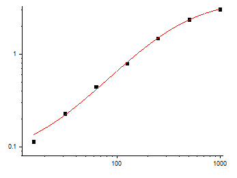
- Warm kit reagents ≥ 30 min RT before opening; protect TMB from light; stop uniformly; read 450 nm promptly; fit 4-PL; run full standard curve per plate (KTE7015's 2-fold serial 15.6–1000 pg/mL, R² ≥ 0.99 per vendor family).
The Bottom Line
TNF-α is the 159-aa (~17.3 kDa monomer, ~51 kDa trimer) type II TM cytokine that sparks the LPS/TLR4 cascade 30 min before IL-1β wakes and 4 h before IL-6 peaks — making it the initiator of septic shock, the driver of CIA joint erosion (the $40B anti-TNF franchise), the early wave of DSS/TNBS colitis, and a bimodal pro-tumor/pro-apoptotic signal in TMEs depending on dose and context. Because it peaks at 1–2 h (LPS) or 6–12 h (CLP) and crashes by 6 h, then runs at < 20 pg/mL baseline, it demands a mouse-specific sandwich ELISA with a single-digit LOD and dilution-linearity that survives the 1:200 pre-dilute of a LPS 1-h bleed. The EliKine™ Mouse TNF-α ELISA Kit — KTE7015 from Abbkine gives you that readout: pre-coated anti-mouse TNF-α capture (trimer-epitope, rejects LT-α/human TNF) → biotin detection → EliKine™ SA–HRP → TMB → 450 nm → pg/mL interpolated, over a 15.6–1000 pg/mL calibrated range with LOD ~8 pg/mL (Intra CV < 8%, Inter CV < 10%), in a ~2.5–3.5 h workflow that scales from a LPS 6-point timecourse to a CIA paw-lavage cohort without chaining


