




Talin-1: The Molecular Clutch of Life – How the Abbkine Human TLN1 ELISA Kit (KTE60318) Deciphers the Code of Cellular Adhesion, Migration, and Metastasis
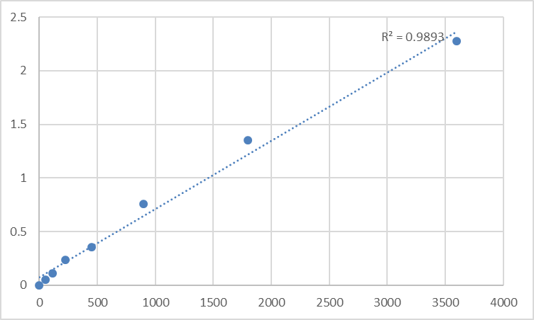
Imagine a microscopic world where a single 270‑kDa protein acts as the central clutch, transmitting mechanical forces between a cell's internal skeleton and its external environment. This protein is Talin‑1 (TLN1), a colossal cytoskeletal adaptor that not only activates integrin receptors but also serves as a primary mechanosensor, translating physical tug‑and‑pull into biochemical signals that dictate cell fate . From immune cell trafficking and wound healing to cancer metastasis and cardiovascular integrity, Talin‑1 sits at the epicenter of cell adhesion signaling . Yet, quantifying this pivotal protein has been a persistent challenge for researchers—Western blots are semi‑quantitative and low‑throughput, while immunofluorescence lacks precise concentration data . The Abbkine Human Talin‑1 (TLN1) ELISA Kit (KTE60318) provides the definitive solution: a high‑sensitivity, sandwich‑format colorimetric assay that accurately measures Talin‑1 in serum, plasma, cell culture supernatants, and other biological fluids with exceptional specificity and reproducibility . Whether you're investigating Talin‑1's role in integrin activation, profiling its expression in metastatic tumors, or screening small‑molecule inhibitors of focal adhesion dynamics, this kit delivers the precision, ease‑of‑use, and scalability needed to advance your research from mechanistic insight to translational impact .
Why Talin‑1 Is the Linchpin of Cellular Mechanics and Disease
Talin‑1 (UniProt Q9Y490) is a 270‑kDa multidomain cytoskeletal protein composed of an N‑terminal FERM (4.1 protein, ezrin, radixin, moesin) head domain and a C‑terminal rod domain comprising 13 helical bundles . Its canonical function is to bridge activated integrins at the plasma membrane to the actin cytoskeleton, forming the core of focal adhesions—the cell's primary force‑transmission hubs . Beyond this structural role, Talin‑1 is a dynamic mechanosensor: applied force unfolds its rod domain, exposing cryptic vinculin‑binding sites that recruit additional actin‑linking proteins, thereby strengthening the adhesion in a positive‑feedback loop . This force‑dependent unfolding enables Talin‑1 to orchestrate focal adhesion turnover, cell spreading, and directional migration—processes fundamental to embryogenesis, immune surveillance, and tissue repair .
In pathology, dysregulated Talin‑1 expression or activity is a hallmark of aggressive cancers. Elevated Talin‑1 promotes integrin‑mediated survival signaling (PI3K/Akt, FAK/Src), enhances invadopodia formation for extracellular matrix degradation, and confers resistance to anoikis (detachment‑induced apoptosis), driving metastasis in prostate, breast, liver, and nasopharyngeal carcinomas . In cardiovascular systems, Talin‑1 mutations or deficiencies disrupt cardiomyocyte costamere organization and vascular endothelial integrity, contributing to dilated cardiomyopathy and aortic dissection . Recent studies even reveal a non‑canonical nuclear role for Talin‑1, where it modulates gene expression by associating with chromatin, providing a direct link between cell‑adhesion events and transcriptional reprogramming . Thus, precise quantification of Talin‑1 protein levels is not merely an assay—it's a window into the mechanical and signaling state of the cell.
Kit Principle: A Robust Sandwich ELISA Designed for Accuracy and Convenience
The Abbkine Human Talin‑1 ELISA Kit employs a classical two‑site immunoenzymometric (sandwich) assay optimized for minimal hands‑on time and maximal reliability . Step 1: A monoclonal anti‑human Talin‑1 antibody is pre‑coated onto the wells of a 96‑well microplate. Step 2: Standards (calibrators) or samples are added; any Talin‑1 present binds to the immobilized capture antibody during a 2‑hour incubation at room temperature. Step 3: After washing away unbound material, a biotin‑conjugated polyclonal anti‑human Talin‑1 detection antibody is added, forming an antibody‑antigen‑antibody “sandwich.” Step 4: Following another wash, Streptavidin‑Horseradish Peroxidase (HRP) conjugate is added, which binds with high affinity to the biotin on the detection antibody. Step 5: A TMB (3,3’,5,5’‑tetramethylbenzidine) substrate is introduced; HRP catalyzes its conversion to a blue chromogen, which turns yellow upon addition of stop solution (sulfuric acid). Step 6: Absorbance is measured at 450 nm (with 570 nm or 630 nm reference optional). The intensity of the yellow color is directly proportional to the amount of Talin‑1 captured in the initial step . Key performance specifications include:
• Detection range: The kit provides a broad dynamic range covering physiologically relevant concentrations in human samples (exact range should be confirmed with the manufacturer's datasheet) .
• Sensitivity (Limit of Detection): High sensitivity enables detection of low‑abundance Talin‑1 in cell‑culture supernatants or early‑disease sera .
• Specificity: The matched antibody pair shows no significant cross‑reactivity with human Talin‑2 or other focal‑adhesion proteins, ensuring accurate quantification even in complex lysates .
• Precision: Intra‑assay and inter‑assay coefficients of variation (CV) are typically <10% and <12%, respectively, guaranteeing reproducible results across operators and plates .
• Sample compatibility: Validated for serum, plasma (EDTA, heparin, citrate), cell‑culture supernatants, and tissue/cell lysates; most samples require only simple dilution before assay .
The entire protocol can be completed in 3–5 hours with less than 2 hours of hands‑on time, making it ideal for medium‑ to high‑throughput screens.
Five Compelling Applications of the Abbkine Talin‑1 ELISA Kit in Cutting‑Edge Research
Research Area Specific Question How KTE60318 Provides the Answer
Cancer Metastasis & Invasion Does Talin‑1 overexpression correlate with invasive potential in triple‑negative breast cancer (TNBC) cell lines or patient‑derived xenografts? Quantify Talin‑1 in cell lysates and conditioned media from migratory vs. non‑migratory populations; correlate with invadopodia activity (gelatin degradation assay) and transwell invasion .
Integrin Signaling & Mechanobiology How do substrate stiffness and geometric confinement alter Talin‑1 expression and focal‑adhesion turnover in mesenchymal stem cells (MSCs)? Measure Talin‑1 in MSCs cultured on soft (0.5 kPa) vs. stiff (50 kPa) hydrogels or micropatterned islands; combine with immunofluorescence for paxillin/vinculin to link protein level to adhesion size/number .
Cardiovascular Remodeling Is plasma Talin‑1 a prognostic biomarker for left‑ventricular hypertrophy or post‑MI remodeling in heart‑failure patients? Profile serial plasma samples from patients undergoing cardiac resynchronization therapy; correlate Talin‑1 levels with echocardiographic parameters (LVEF, LV mass) and NT‑proBNP .
Immunology & Leukocyte Trafficking Does Talin‑1 knockdown impair T‑cell adhesion and trans‑endothelial migration in models of inflammation or autoimmunity? Compare Talin‑1 in wild‑type vs. Talin‑1‑KO T‑cell lysates after stimulation on ICAM‑1/VCAM‑1 coats; link to flow‑based adhesion assays under shear stress .
Drug Discovery & Target Validation Can a small‑molecule inhibitor of the Talin‑1–integrin β‑tail interaction reduce Talin‑1‑mediated signaling in cancer cells? Use the ELISA to measure Talin‑1 in cell lysates after compound treatment as a pharmacodynamic readout; combine with FAK/paxillin phosphorylation assays to confirm pathway inhibition .
Step‑by‑Step Workflow: From Sample to Data in One Afternoon
① Sample Preparation
• For cell lysates, lyse cells in RIPA buffer supplemented with protease/phosphatase inhibitors; centrifuge at 12,000 × g for 15 min at 4°C to remove debris.
• For serum/plasma, collect blood in EDTA tubes, centrifuge at 1,000–2,000 × g for 15 min within 30 min of collection; aliquot and store at –80°C.
• For cell‑culture supernatants, centrifuge at 500 × g for 5 min to remove cells and debris.
• Avoid repeated freeze‑thaw cycles (>3 cycles may degrade Talin‑1); thaw samples on ice and dilute if necessary using the provided assay buffer.
② Reagent Preparation
• Equilibrate all components to room temperature (18–25°C) for 30 min before use.
• Reconstitute the lyophilized Talin‑1 standard with the provided diluent to generate the stock solution.
• Prepare a 7‑point standard curve via serial dilution (e.g., 1:2 dilutions) covering the kit's dynamic range.
• Prepare Working Detection Antibody by diluting the biotin‑conjugated antibody per instructions.
• Prepare Streptavidin‑HRP Working Solution by diluting the conjugate as directed.
• Prepare 1× Wash Buffer by diluting the 20× concentrate with distilled water.
③ Assay Procedure
• Add 100 µL of standard or sample to appropriate wells (duplicate/triplicate recommended).
• Cover with plate sealer and incubate 2 hours at room temperature with gentle shaking.
• Aspirate and wash each well 4 times with 300 µL Wash Buffer (soak 30 sec per wash).
• Add 100 µL of Working Detection Antibody; incubate 1 hour at room temperature.
• Wash as in step 3.
• Add 100 µL of Streptavidin‑HRP Working Solution; incubate 45 minutes at room temperature.
• Wash as in step 3.
• Add 100 µL of TMB Substrate; incubate 15–30 minutes at room temperature in the dark.
• Add 50 µL of Stop Solution; read absorbance at 450 nm within 30 min.
④ Data Analysis
• Subtract the average absorbance of the blank (zero standard) from all standards and samples.
• Plot the standard curve (absorbance vs. concentration) using a 4‑parameter logistic (4PL) or log‑log curve‑fit.
• Interpolate sample concentrations from the curve; multiply by dilution factor if applicable.
• For values outside the range, re‑assay at an appropriate dilution.
⑤ Quality Control
• Include a positive control (recombinant human Talin‑1) and a pooled human serum/lysate sample in each run.
• Acceptable criteria: standard‑curve R² >0.99, CV of replicates <15%, spike‑recovery 80–120%.
How the Abbkine Kit Stacks Up Against Other Talin‑1 Detection Methods
Method Principle Sensitivity Time per 96 Samples Throughput Best For
Abbkine Sandwich ELISA (KTE60318) Colorimetric sandwich ELISA with pre‑coated plate, biotin‑streptavidin amplification. High (pg/mL‑ng/mL range) 3–5 hours High (96‑well plate) Routine quantification in multiple sample types; medium‑ to high‑throughput screens.
Western Blot SDS‑PAGE separation, transfer, anti‑Talin‑1 antibody detection. Moderate (ng‑µg range, semi‑quantitative) 1–2 days Low (10–20 samples/gel) Confirming Talin‑1 molecular weight (~270 kDa) and detecting proteolytic fragments.
Immunofluorescence (IF) Antibody staining followed by fluorescence microscopy. Qualitative/Low quantitative 1–2 days Low (manual imaging) Subcellular localization at focal adhesions; co‑localization with integrins/vinculin.
Proximity Ligation Assay (PLA) Amplified fluorescence signal when two antibodies are in close proximity. High (single‑molecule detection) 1–2 days + imaging Low Detecting Talin‑1–integrin or Talin‑1–vinculin interactions in situ.
Mass Spectrometry (MS) LC‑MS/MS quantification using stable‑isotope‑labeled peptides. Very High (attomole level) 2–3 days + data analysis Medium (multiplexed) Absolute quantification; post‑translational modification (phosphorylation) mapping.
SimpleStep ELISA (e.g., Abcam) Single‑wash, one‑step incubation technology. Comparable (0.044–0.22 ng/mL) 1.5 hours High (96‑well plate) Ultra‑fast screening when time is critical.
The Abbkine kit strikes an optimal balance between sensitivity, throughput, and cost, offering excellent specificity with a traditional sandwich format that is familiar to most lab personnel. It is more quantitative than Western blot, faster than IF/PLA, and more accessible than MS for most academic and industrial labs.
Troubleshooting Guide: Ensuring Reliable Talin‑1 Quantification
Issue Possible Cause Solution
High background in blank wells Contaminated wash buffer (bacterial growth); incomplete washing (residual HRP); plate sealer reused (carryover). Use fresh wash buffer prepared with Milli‑Q water; ensure 30‑sec soak per wash; use new plate sealers for each incubation.
Poor standard‑curve linearity (R² <0.98) Improper serial dilution (pipetting error); standard reconstituted incorrectly (not mixed thoroughly); incubation temperature fluctuations. Use calibrated pipettes; vortex standard stock for 30 sec after reconstitution; perform all incubations in a temperature‑controlled incubator/shaker.
Low signal in cell lysates Insufficient protein concentration (Talin‑1 below detection limit); lysis buffer incompatible (strong detergents interfere). Concentrate lysates using 10‑kDa centrifugal filters; ensure lysis buffer is compatible (avoid SDS >0.1%); optimize protein loading (10–50 µg/well).
High intra‑assay variability (CV >15%) Inconsistent pipetting (especially viscous sera/lysates); bubble formation during reagent addition; edge effects in plate. Use reverse‑pipetting technique for viscous samples; tap plate gently after each addition; avoid using outer wells or pre‑condition plate with assay buffer.
Signal saturation at high concentrations Sample Talin‑1 concentration above kit's range (e.g., concentrated tumor lysates). Pre‑dilute samples 1:10–1:100 in assay buffer and re‑assay; confirm dilution linearity via spike‑and‑recovery.
Discrepancy between duplicate wells Inadequate mixing of samples before loading; uneven washing (clogged washer needles). Vortex samples briefly before loading; check washer nozzles for obstruction; ensure plate is leveled during washing.
Best Practices for Publication‑Ready Talin‑1 Data
Practice Rationale & Implementation
Standardize sample collection & processing For cell experiments, lyse cells at consistent confluence (e.g., 80–90%) and normalize to total protein (BCA assay). For clinical samples, collect serum/plasma in EDTA tubes (inhibits calcium‑dependent proteases); process within 2 hours; store at –80°C in single‑use aliquots.
Include appropriate controls Always run a blank (zero standard), a kit‑provided positive control, and a matrix‑matched control (e.g., pooled normal serum or untransfected cell lysate) in each plate.
Validate dilution linearity & spike recovery For each new sample type (e.g., ascites, synovial fluid), perform a spike‑and‑recovery experiment using recombinant Talin‑1; acceptable recovery is 80–120%.
Use a 4‑parameter logistic (4PL) curve fit The 4PL model accurately captures the sigmoidal ELISA standard curve; avoid linear regression, which underestimates high and low concentrations.
Benchmark against an orthogonal method Periodically compare ELISA results with Western blot densitometry or MS‑based quantification to ensure correlation (expect R² >0.90).
Document kit lot numbers & storage conditions Record the kit lot number, standard reconstitution date, and storage temperature; this is essential for publication and ensures reproducibility across experiments.
Randomize sample placement To avoid plate‑position bias, randomize samples across the plate rather than grouping all controls or treatments together.
From Basic Science to Clinical Translation: Where Talin‑1 Quantification Makes an Impact
① Oncology biomarker discovery
The kit enables serum Talin‑1 profiling in longitudinal cohorts of breast, prostate, or colorectal cancer patients, correlating levels with metastasis‑free survival, response to integrin‑targeted therapies (e.g., cilengitide), or resistance to chemotherapy .
② Cardiovascular disease research
Researchers can quantify Talin‑1 in plasma from patients with aortic aneurysms or heart failure, exploring its utility as a prognostic marker for ventricular remodeling or vascular dissection risk .
③ Immunology & autoimmunity
The assay facilitates measurement of Talin‑1 in T‑cell or neutrophil lysates from autoimmune models (e.g., rheumatoid arthritis, multiple sclerosis), linking Talin‑1 expression to integrin activation status and leukocyte adhesion/migration .
④ Drug screening & target engagement
Pharma labs use the kit as a pharmacodynamic readout in high‑throughput screens for Talin‑1–integrin disruptors or allosteric inhibitors of Talin‑1 unfolding, accelerating lead‑optimization cycles.
⑤ Stem‑cell & tissue engineering
By monitoring Talin‑1 levels in MSC‑derived extracellular vesicles or engineered tissue constructs, scientists can assess mechanical maturation and integrin signaling activity as quality‑control metrics.
A Ready‑to‑Use Methods Section for Your Manuscript
Talin‑1 protein concentration was quantified using the Abbkine Human Talin‑1 (TLN1) ELISA Kit (KTE60318) according to the manufacturer's instructions. Briefly, cell lysates (20 µg total protein) or serum samples (diluted 1:10 in assay buffer) were added to anti‑Talin‑1 pre‑coated 96‑well plates and incubated for 2 h at room temperature with shaking. After washing, biotinylated detection antibody was added for 1 h, followed by Streptavidin‑HRP conjugate for 45 min. TMB substrate was added for 20 min, the reaction stopped with sulfuric acid, and absorbance measured at 450 nm on a microplate reader (BioTek Synergy H1). A four‑parameter logistic (4PL) standard curve was used to interpolate sample concentrations. All samples were assayed in duplicate, and the mean intra‑assay coefficient of variation (CV) was <10%. Talin‑1 levels are expressed as ng/mL (serum/plasma) or ng/mg total protein (lysates).
Why the Abbkine Human Talin‑1 ELISA Kit (KTE60318) Is the Go‑To Tool for Adhesion Biology
① It captures Talin‑1's multifaceted biology – by providing precise, quantitative data across diverse sample types, the kit enables researchers to dissect Talin‑1's roles not only in focal adhesion mechanics but also in nuclear signaling, immune‑cell trafficking, and cancer metastasis.
② It accelerates translational workflows – with a 3–5‑hour protocol and pre‑coated plates, core facilities can process hundreds of clinical or preclinical samples per week, supporting biomarker validation studies or compound screens.
③ It conserves precious samples – requiring only 100 µL of serum/plasma or 20–50 µg of lysate protein, the kit allows multiplexed analysis of Talin‑1 alongside other adhesion molecules (FAK, paxillin, vinculin) from the same aliquot—critical for limited biobank specimens or pediatric samples.
④ It delivers publication‑grade reproducibility – the matched antibody pair and optimized buffers yield intra‑assay CVs <10% and inter‑assay CVs <12%, meeting the stringent reproducibility standards of journals like Journal of Cell Biology and Cancer Research.
⑤ It's backed by Abbkine's quality guarantee – each lot is QC‑tested for sensitivity, specificity, and spike‑recovery, and the company provides detailed technical support, protocol optimization guides, and a 30‑day satisfaction guarantee.
Ready to unlock the secrets of cellular adhesion and mechanotransduction? The Abbkine Human Talin‑1 (TLN1) ELISA Kit (KTE60318) delivers exceptional sensitivity, specificity, and ease‑of‑use—whether you're mapping Talin‑1's role in cancer progression, decoding integrin signaling networks, or screening next‑generation anti‑metastatic therapeutics. With pre‑coated plates, ready‑to‑use reagents, and results in one afternoon, it's the turnkey solution for precision Talin‑1 quantification.
🔗 Product reference: KTE60318 (Abbkine) – https://www.abbkine.com/product/human-talin-1-tln1-elisa-kit-kte60318/
(For research use only. Not for diagnostic or therapeutic procedures. Store at 2–8°C protected from light; stable for 12 months.)





