




The Plasma Membrane Sentinel: Na+/K+-ATPase α1 Rabbit Polyclonal Antibody (ABL1141)
A first-year PhD student in a neuroscience lab recently confessed something that would resonate with every electrophysiologist who ever lived. She had spent six months characterizing a novel KCNT1 channel mutation linked to severe nocturnal frontal lobe epilepsy, running patch-clamp recordings that showed clear hyperexcitability, and building a compelling case for a gain-of-function mechanism. But when the time came to normalize her western blot data for the companion molecular biology experiments, she reached for β-actin. The problem was not that β-actin failed to produce a band. The problem was that her treatment condition—a pharmacological intervention targeting membrane excitability—altered the ratio of membrane to cytosolic protein in her lysates. Every lane on her SDS-PAGE gel contained a different proportion of plasma membrane protein, and β-actin, a cytosolic protein that does not inhabit the compartment being studied, was reporting loading for a subcellular fraction it did not represent. The normalization was systematically biased, and the error propagated through every quantification that followed.
Na+/K+-ATPase α1 does not have this problem. It is an integral plasma membrane protein—the catalytic subunit of the sodium-potassium pump that establishes and maintains the electrochemical gradients of Na⁺ and K⁺ ions across the plasma membrane of every animal cell. These gradients are essential for osmoregulation, for sodium-coupled transport of a variety of organic and inorganic molecules, and for electrical excitability of nerve and muscle. Unlike diffusible cytoplasmic markers whose abundance in a membrane-enriched fraction depends on the efficiency of the enrichment protocol, Na+/K+-ATPase α1 resides where the biological action occurs—at the membrane itself. When a researcher fractionates cellular compartments, α1 follows the plasma membrane fraction with stoichiometric fidelity. When a treatment alters membrane protein trafficking, α1 reports it. When a reviewer asks whether the loading control actually reflects the compartment being analyzed, the answer is yes.
Abbkine's Na+/K+-ATPase α1 Rabbit Polyclonal Antibody (ABL1141) enters this analytical landscape with specifications that reward a close reading of the datasheet. The antibody was raised in rabbit against a synthesized peptide derived from human Na+/K+-ATPase α1 around the non-phosphorylation site of serine 16, a region selected to ensure that the phosphorylation state of the enzyme—which fluctuates dynamically under physiological regulation—does not alter antibody binding affinity. The antiserum was then affinity-purified by affinity-chromatography using the epitope-specific immunogen, enriching the final product for antibodies that recognize the S16 region specifically while depleting those that bind other epitopes on the α1 subunit or cross-react with the structurally homologous α2, α3, and α4 isoforms. This is not a crude antiserum. It is an affinity-purified polyclonal whose epitope specificity has been narrowed to a defined region of the α1 N-terminal domain.
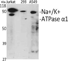
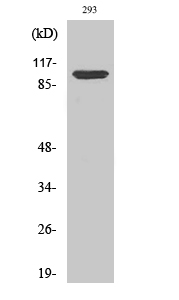
The epitope selection strategy deserves emphasis because it determines what the user actually sees on the membrane. Na+/K+-ATPase α1 runs at approximately 112 kDa on reducing SDS-PAGE, a molecular weight well separated from the ~100 kDa region where the α2 and α3 isoforms migrate and far removed from the ~55 kDa β-subunit. The product page confirms that the antibody detects endogenous levels of total Na+/K+-ATPase α1 protein and produces a clean single band at 112 kDa in western blot analysis of various cells and 293 cells at a dilution of 1:1000. The band is sharp, the background is clean, and the signal intensity reflects α1 abundance specifically rather than a pooled Na+/K+-ATPase isoform summation—a specificity profile that distinguishes ABL1141 from generic pan-α antibodies that cannot discriminate between the four catalytic subunit isoforms with distinct tissue distributions and regulatory properties.
Species reactivity spans human, mouse, and rat, the three mammalian species that account for the overwhelming majority of cell biology, neuroscience, and preclinical pharmacology publications. The S16 non-phosphorylation site is highly conserved across these three species, and the antibody's reactivity reflects that conservation. For a core facility processing western blots from human HEK293 lysates, mouse brain homogenates, and rat hippocampal neuron cultures in a single day, this cross-reactivity eliminates the logistical burden of maintaining separate membrane loading control antibodies for each organism.
Applications extend across four core immunoassay modalities: western blot at a suggested starting dilution of 1:500–1:2000, immunohistochemistry on paraffin-embedded tissue at 1:100–1:300, immunofluorescence at 1:200–1:1000, and ELISA at 1:10000. The dilution ranges reflect genuine optimization rather than marketing optimism—western blot data on the product page show clean performance at 1:1000, and the broader recommended range accommodates the variability in endogenous α1 expression across different tissue types. The IF and IHC-P validations mean the same antibody can detect α1 in its native membrane context, revealing the spatial distribution of the sodium pump in tissue sections and cultured cells without requiring separate antibodies for imaging versus blotting applications.
Formulation and storage specifications reward a practical reading. The antibody is supplied as a liquid solution at 1 mg/mL in PBS containing 50% glycerol, 0.5% BSA, and 0.02% sodium azide as preservative. The 50% glycerol depresses the freezing point, preventing ice crystal formation that denatures immunoglobulin protein during -20°C storage. The 0.5% BSA serves as a carrier protein that stabilizes the antibody against surface adsorption and denaturation during freeze-thaw cycling—a formulation detail that extends the functional shelf life beyond what glycerol alone can provide. Storage instructions specify one-year stability at -20°C from the date of shipment, with centrifugation of the original vial after thawing and prior to cap removal recommended for maximum product recovery, and aliquoting advised to avoid repeated freezing and thawing. Shipping is on gel packs with blue ice. The product is for research use only and is not intended for use in human or clinical diagnosis. Available in 50 µL and 200 µL vial sizes, with a 30 µL option accessible through certain procurement platforms.
The publication record for ABL1141 currently includes six citations in peer-reviewed literature, and the identity of these publications reveals more about the antibody's performance than the number alone. One study published in JCI Insight (IF 8.2) deployed the antibody while characterizing a novel small molecule AdipoR2 agonist that ameliorates experimental hepatic steatosis in hamsters and mice—a metabolic disease context requiring membrane protein normalization in tissue samples where lipid overload could alter membrane composition and protein extraction efficiency. A second publication in the British Journal of Pharmacology (IF 7.7) used ABL1141 during the investigation of carvedilol inhibition of neuronal hyperexcitability caused by epilepsy-associated KCNT1 mutations—precisely the kind of study where a membrane-resident loading control is analytically essential because the experimental treatment alters membrane excitability itself. A third study, published in Cell Death & Disease, identified the acid-sensitive site critical for chloral hydrate activation of the proton-activated chloride channel. A fourth publication in Cell Reports Medicine and a fifth in the Journal of Experimental & Clinical Cancer Research (IF, as listed in the product page links) further extend the validated application range of ABL1141 into translational biomedicine. An additional study, detailed on the Abbkine news page, employed the antibody to elucidate the role of N-myristoylation in the excessive membrane localization of PD-L1 in hypoxic cancers and to develop a novel NMT1 inhibitor for combination with immune checkpoint blockade therapy.
For the electrophysiologist characterizing ion channel mutations in transfected cells, the cancer biologist quantifying membrane protein trafficking, and the metabolism researcher studying hepatic sodium pump regulation in steatosis models, the band at 112 kDa on the membrane tells a truth that cytoplasmic loading controls cannot access: it reports the protein content of the membrane fraction, and it is unaffected by the phosphorylation state of the target epitope.
Explore full specifications, view representative images, and place your order here: https://www.abbkine.com/product/na-k-atpase-%ce%b11-rabbit-polyclonal-antibody-abl1141/





