


The 30-kDa Ring at the Heart of the Mitochondrion: Why Quantifying Prohibitin (PHB1) Actually Matters — And How KTE61222 Turns It Into a Real Number
If you've ever run a subcellular fractionation and reached for "Prohibitin" as your inner mitochondrial membrane (IMM) loading control, you already know the protein — but what most people forget is that PHB isn't just a convenient marker band at ~30 kDa; it's a structurally essential, evolutionarily frozen scaffold that holds the respiratory apparatus together, gates mitophagy, and moonlights in the nucleus as a transcriptional co-regulator. Officially Prohibitin (PHB / PHB1, UniProt: P35232, Gene ID: 5245), this 272-aa, ~29.8 kDa protein assembles with its sibling PHB2 into large ring-shaped ~1 MDa complexes in the IMM that act as a chaperone/stability scaffold for respiratory chain proteins, maintain cristae morphology, regulate cardiolipin remodeling, and serve as the OMA1 proteolytic checkpoint interface. The catch? In many labs, PHB still lives as a qualitative Western blot band — "it's there, move on" — when the actual experiment (mitochondrial biogenesis, metabolic stress, apoptosis, cancer metabolism, or neurodegeneration) needs it quantified as a reproducible, plate-readable concentration. The Human Prohibitin (PHB) ELISA Kit (KTE61222) from Abbkine exists to close that gap: a quantitative two-site sandwich ELISA that converts the most famous mitochondrial marker in biology into ng/mL (or pg/mL) with a standard curve, so your fractions, treatments, and disease-model lysates stop relying on eyeball densitometry.
PHB / PHB1 in One Clean Pass: Not a "Tumor Suppressor" in the Way You Were Taught
The human PHB gene sits at 17q21.33 — the same BRCA1 neighborhood that guaranteed it attention early on — and for years it was marketed as a negative regulator of cell proliferation / tumor suppressor because over-expression slowed growth. The field has since cleaned this up: the anti-proliferative punch actually lives in the 3′-UTR (regulatory RNAs, miRNA-binding elements), not the PHB polypeptide itself. The protein's real job description is far more architectural:
Compartment What PHB (as PHB1–PHB2 ring complex) Actually Does
Mitochondrial IMM Scaffolds/protects respiratory complexes, stabilizes nucleoids, organizes cristae via cardiolipin contacts, regulates OMA1 turnover, functions as a mitophagy receptor (LC3-II interaction upon depolarization)
Nucleus Transcriptional coregulator — enhances p53 promoter binding, represses E2F1, modulates ER/androgen receptor activity via HDAC recruitment
Plasma membrane / signaling endosomes Associates with signaling complexes (CD86 cytoplasmic tail, ARFGEF3, SPHK2, IGFBP6) — less a "membrane structural protein" than a signaling-integration node
Translation: PHB is one of those rare proteins that is both the most boringly ubiquitous mitochondrial marker and one of the most mechanistically connected hubs between energy homeostasis, quality control, and transcriptional control. When you measure it, you're not measuring fluff — you're measuring an organizational backbone.
Why a Sandwich ELISA — And Why "Just Probe It on a Gel" Leaves Money on the Table
PHB is hydrophobic, ~30 kDa, and IMM-integral, which means:
• If you under-centrifuge or over-dilute your mitochondrial fraction, the "band" gets faint not because mitochondria vanished but because your recovery did.
• Densitometry across gels with variable transfer efficiency at 30 kDa (near the stacking/gel-transition zone) accumulates error fast.
• Any experiment that needs ≥20 samples × replicates × fractions will burn days on transfers and still have "is this linear?" anxiety.
The KTE61222 sandwich ELISA dodges this with the classic, high-specificity architecture:
- Microplate pre-coated with an anti-PHB capture antibody.
- Standards (recombinant human PHB) and samples (serum, plasma, tissue homogenates, cell lysates, cell culture supernatants/other biological fluids) added → PHB binds.
- Wash → biotinylated anti-PHB detection antibody (different epitope) → Streptavidin–HRP.
- TMB → color ∝ bound PHB → stop → read 450 nm → interpolate from the PHB standard curve.
From distributor-matched references, the operating specs cluster around:
Parameter Typical KTE61222-class Specification
Target Human Prohibitin / PHB1 (UniProt P35232, 272 aa, ~29.8 kDa)
Assay type Quantitative sandwich ELISA, pre-coated capture
Detection Biotin-Ab → SA‑HRP → TMB, 450 nm
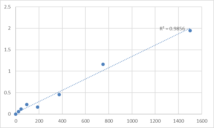
Dynamic range ~0.31 – 20 ng/mL (7‑point standard)
Sensitivity / LOD 0.1 – 0.2 ng/mL (6–115 pg/mL depending on format/sensitivity definition)
Intra-Assay CV < 8–10%
Inter-Assay CV < 10–12%
Specificity No significant cross‑reactivity with PHB2/other analogues
Samples Serum, plasma, tissue homogenates, cell lysates, culture supernatants, other biological fluids
Assay time ~3–5 hours
(As always, anchor your Methods to the shipped lot‑specific certificate of analysis.)
Where Quantifying PHB Protein Moves the Needle
- Subcellular fractionation & mitochondrial enrichment QC
This is the single most common "hidden use": you've spun a differential centrifugation (600 ×g → 10,000 ×g pellet = PM + mitochondria-enriched, etc.), and you want a quantitative anchor that says "my IMM load is actually X ng/mg, not 'the PHB lane looks similar.'" ELISA lets you normalize across preparations and detect subtle losses that wreck respiratory assays.
- Mitochondrial biogenesis / dynamics (PGC‑1α, PPARγ, ERRα programs)
If your treatment drives mitochondrial proliferation (exercise, resveratrol, bezafibrate, thyroid hormone, BMP7 in podocytes), total PHB is one of the cleanest proxy reads for "how much IMM mass did I actually build?" — especially when paired with VDAC/porin (outer membrane) and COX IV/TFAM as complementary markers.
- Metabolic stress, I/R injury & oxidative phosphorylation screens
Ischemia–reperfusion, rotenone/antimycin, oligomycin, and nutrient starvation all stress the IMM scaffold. PHB can shift in solubility or associate with aggregates under proteotoxic stress — and quantifying it (rather than assuming it's constant) prevents you from misreading your OXPHOS enzyme activity data.
- Cancer metabolism & apoptosis resistance
PHB is up-regulated in several tumors and can protect against drug-induced apoptosis by stabilizing mitochondrial integrity. CRISPR knockouts of PHB1 sensitize cells to mitochondrial stressors — and your validation should be % PHB protein remaining ± SEM from a calibrated curve, normalized to total protein, not a "lighter vs. darker" band.
- Neurodegeneration & mitophagy
Because the PHB1–PHB2 complex is a mitophagy receptor (LC3-II interaction post-depolarization), tracking PHB levels across temporal Parkinson's models (MPTP/α-syn/PINK1-PRKN) or aging panels gives you a structural readout of the IMM's "handover machinery" — best expressed as ng PHB / mg total protein (BCA) so the mitochondrial pool is comparable across conditions.
A Minimal Prep Note So Your 450 nm Reflects Mitochondria, Not Debris
PHB is integral membrane — treat it that way:
• Homogenize cold in 250 mM sucrose / 10 mM Tris, pH 7.4 + protease inhibitors; do your differential spins (nuclei → crude mito @ ~10,000 ×g → (optional) purify on Percoll/sucrose gradient).
• For whole-cell/whole-tissue work, lyse in RIPA or 0.5–1% Triton X-100 + 150 mM NaCl, pH 7.4, clarify ~16,000 ×g, keep supernatant cold.
• BCA the same lysate → report ng PHB / mg total protein.
• Warm all kit reagents ≥ 30 min RT before opening, protect TMB from light, stop uniformly, read 450 nm promptly, and run the full standard curve on every plate — membrane-protein recovery is exactly why "Tuesday's curve" can't save "Thursday's plate."
The Bottom Line
Prohibitin (PHB1) is the ~30 kDa ring that quietly holds your cristae — and your respiratory chain — together, then shows up in the nucleus to whisper to p53 and E2F1, and even waves at the autophagosome when mitochondria break bad. Measuring it deserves more than "it's the loading control, next lane." The Human Prohibitin (PHB) ELISA Kit — KTE61222 from Abbkine gives you a pre-coated sandwich ELISA that turns this mitochondrial cornerstone into a calibrated variable: capture → biotin detection → HRP–TMB → 450 nm → ng/mL, over a ~0.31–20 ng/mL range, in a ~3–5 hour workflow that scales across fractions, treatments, and cohorts without chaining you to a gel rig.
Product Reference: KTE61222 – Human Prohibitin (PHB) ELISA Kit
Learn more and order: https://www.abbkine.com/product/human-prohibitin-phb-elisa-kit-kte61222/
(For Research Use Only; not for diagnostic procedures in humans.)


