


Not PlGF, Not VEGF: Why the “Other” PIGF—the GPI‑Anchor Assembly Factor—Deserves Its Own ELISA, and How Abbkine’s KTE61212 Finally Lets You Quantify It
If your lab works anywhere near hematopoiesis, paroxysmal nocturnal hemoglobinuria (PNH), or the crowded world of surface‑protein anchors, you’ve almost certainly mistyped “PIGF” into a search bar and landed on the wrong molecule. Popular usage has hijacked the acronym: in most PubMed-adjacent conversations, PIGF / PGF means Placenta Growth Factor, a VEGF‑family secreted cytokine. But the protein on this page is the other PIGF—Phosphatidylinositol‑Glycan biosynthesis Class F protein, gene symbol PIGF (UniProt: Q07326, also called PIG‑F / GPI11), a ~25–27 kDa factor that lives in the ER/nucleoplasmic GPI‑anchor biosynthesis pathway and helps shepherd the pre‑GPI intermediate through the assembly line that ultimately tethers CD55, CD59, CD157/BST1, alkaline phosphatase, and hundreds of other proteins to the outer leaflet of the plasma membrane. The Human Phosphatidylinositol‑glycan biosynthesis class F protein (PIGF) ELISA Kit (KTE61212) from Abbkine exists to do one simple, powerful thing: turn this historically “invisible” ER‑resident assembly factor into a calibrated, plate‑readable concentration (pg/mL) you can normalize, repeat, and—crucially—correlate with GPI‑anchor integrity, hematopoietic stem/progenitor phenotypes, and PNH‑clone dynamics.
PIGF (PIG‑F) in a Single Sentence: The Gateway Factor You Rarely Measure but Can’t Afford to Ignore
The GPI anchor is not a protein—it’s a phosphatidylinositol‑based glycolipid built stepwise in the ER membrane; the finished anchor is then transamidated onto target proteins bearing the signature C‑terminal GPI‑attachment signal (ω‑site). PIGF participates in the middle/later phase of this assembly: it associates with the GPI transamidase complex (GPI‑T: GPAA1–PIGK–PIGS–PIGT–PIGU/PIGW) and is implicated in ethanolamine‑phosphate addition steps and in coordinating the hand‑off of pre‑GPI intermediates so the transamidase can do its job.
The clinical punchline? The canonical acquired GPI‑defect disease is PNH, where an X‑linked PIGA somatic mutation in a hematopoietic stem cell knocks out the very first step (GlcN‑acyl‑PI synthesis), producing a PNH clone whose progeny lack all GPI‑anchored proteins (CD55⁻ CD59⁻ …) and become complement‑sensitive, thrombosis‑prone, and anemia‑driving. While PIGF itself is not the PNH target gene, its protein level is a readout of whether the GPI pathway “machinery” is present and intact—which is exactly why you sometimes need more than a flow‑cytometry CD55/CD59 scatterplot to understand why an anchor pathway is failing (nutritional ER stress, riboflavin/FAD‑linked cofactor issues, off‑target drug toxicity, CRISPR multi‑gene interactions).
Why a Sandwich ELISA for PIGF—And Why It’s Not Just “Another Housekeeper”
PIGF is not secreted, not abundant like a cytokine, and mostly cytosolic/nucleoplasmic with ER proximity—so your “signal” lives in cells/tissues, not in a neat supernatant band you can skim. That creates three practical pains:
- It hides in whole‑cell or ER‑enriched lysates—if you only look at a surface stain, you never touch PIGF.
- Antibody specificity matters doubly: some databases lump “PIGF” searches into PlGF/VEGF unless you gate on gene ID 5281 and UniProt Q07326.
- Replicates and treatment panels (vitamin B2/B12/folate‑linked cofactor shifts, ER‑stress inducers, differentiation timecourses) demand a plate‑based number, not “band looks fainter.”
The KTE61212 kit uses the field‑standard two‑site sandwich ELISA:
• Pre‑coated anti‑PIGF capture antibody on 96‑well plates
• Standards (recombinant human PIGF) + samples → PIGF binds
• Wash → biotinylated anti‑PIGF detection antibody (different epitope)
• Streptavidin–HRP → TMB → 450 nm → interpolate from the PIGF standard curve
The widely quoted performance envelope you’ll cite:
Parameter Typical KTE61212‑class specification
Target Human PIGF / Phosphatidylinositol‑glycan biosynthesis class F protein (Gene 5281, UniProt Q07326)
Aliases PIG‑F, GPI11 homolog
Format 96‑well sandwich ELISA, pre‑coated capture
Detection Biotin‑Ab → SA‑HRP → TMB, 450 nm
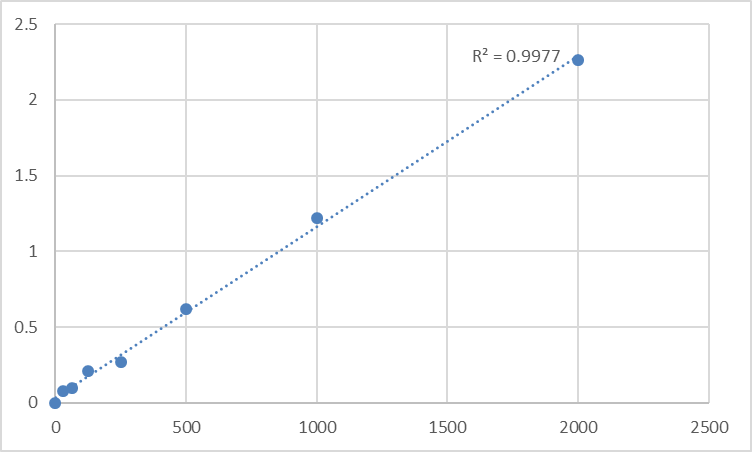
Range 31.2 – 2000 pg/mL (reconstituted standard series)
Sensitivity / LOD ~15–31 pg/mL
Samples Cell culture supernatants*, serum, plasma, tissue homogenates, other biological fluids
Assay time ~3–5 h (depends on user/step timing)
Status For Research Use Only — NOT for diagnostic procedures
*PIGF is not a secreted cytokine; any “supernatant” signal typically reflects cell lysis/debris or vesicle/microsome shedding—clarify by centrifugation and treat conservatively.
Where Quantifying PIGF Actually Adds Leverage
- PNH & GPI‑anchor deficiency research (the strongest “why”)
Flow cytometry shows you CD55/CD59 loss on the surface; a PIGF ELISA on bone‑marrow/target‑cell lysates shows you whether the assembly line proteins are even there. If PIGF drops alongside GPAA1/PIGS/PIGT in a model, you’re looking at a machinery‑wide collapse, not just a PIGA singular hit—and that distinction guides rescue strategy (AAV‑PIGA vs. broader ER cofactor/nutrition correction).
- Hematopoietic stem/progenitor culture & HSC‑expansion bioprocessing
Ex vivo HSC expansion can stress the ER (hypoxia↔re‑oxygenation, cytokine cocktails, small‑molecule “niche” drugs). Tracking PIGF (and CD59 re‑expression after purification) is a quality‑control readout that your anchor proteome is intact before you infuse/re‑engraft.
- ER‑stress, UPR, and riboflavin/FAD‑linked cofactor models
GPI‑anchor assembly is lipid‑dependent and ER‑quality‑control sensitive; PIGF protein levels (normalized to β‑actin/GAPDH/β‑tubulin or total protein by BCA) give you a lightweight proxy for whether your ER‑stressor is spilling into anchor biosynthesis.
- CRISPR/RNAi screens on the GPI pathway
Knocking PIGF (or GPAA1, PIGS, PIGT, PIGA)? Don’t rely on “CD59 looks dimmer” alone—report % PIGF protein remaining ± SEM from a calibrated curve, and if possible, co‑show FLAER‑ALEXA binding + CD59 surface MFI so the clone’s GPI status is defined both inside (assembly factor) and outside (anchor presence).
- Lymphoid/AML lines with spontaneous GPI‑anchor variants
Some laboratories select GPI‑negative sublines (e.g., based on aerolysin/Proaerolysin resistance) to dissect anchor genetics; PIGF ELISA is the fast “machinery check” across clones without dedicating a week to subcellular fraction Westerns.
A Clean Prep/Workflow You Can Paste Into a Protocol Book
For cultured cells / tissue (the real signal source):
- Lyse cold in 50–100 mM Tris, pH 7.4, 150 mM NaCl, 0.5–1% Triton X‑100/NP‑40 + protease inhibitors.
- Clarify 12,000–16,000 ×g, 4°C, 15 min (keep supernatant—this is where soluble/nucleoplasmic PIGF + loose membrane‑associated PIGF ends up).
- BCA the same final lysate → express as pg PIGF / mg total protein.
- Warm all kit reagents ≥30 min to RT before opening; protect TMB from light; stop uniformly; read 450 nm promptly; fit a 4‑PL; back‑calculate dilutions.
- Run the full standard curve on every plate—ER‑resident factors are unforgiving when prep drifts.
Bottom Line
PIGF (PIG‑F) is not the flashy growth factor everyone names at conferences; it’s the background‑essential assembler that decides whether CD55, CD59, and the entire GPI‑anchored surfaceome even exist. If your project touches PNH, HSC quality control, ER‑stress phenotypes, or GPI‑anchor pathway genetics, you need a way to measure it that isn’t “guess the band.” The Human Phosphatidylinositol‑glycan biosynthesis class F protein (PIGF) ELISA Kit — KTE61212 from Abbkine gives you that: pre‑coated capture → biotin detection → HRP–TMB → 450 nm, with a 31.2–2000 pg/mL working range and ~15–31 pg/mL sensitivity, in a ~3–5 h workflow that scales across clones, conditions, and timepoints.
Product Reference: KTE61212 – Human Phosphatidylinositol‑glycan biosynthesis class F protein (PIGF) ELISA Kit
Learn more and order: https://www.abbkine.com/product/human-phosphatidylinositol-glycan-biosynthesis-class-f-protein-pigf-elisa-kit-kte61212/
(For Research Use Only; not for diagnostic procedures in humans.)


