




The 14-Citation Signal Nobody Talks About—And the Color Reaction Behind It
Any metabolite whose concentration you can measure with a $15 drugstore meter seems, at first glance, too pedestrian to deserve space in a peer-reviewed methods section. Glucose is the biochemical water cooler—everyone draws from it, nobody writes poems about it. But that ubiquity is precisely what makes glucose quantification the most quietly dangerous assay in a metabolism laboratory. A 20% systematic error in a glucose measurement propagates through every downstream calculation that depends on it: glycolytic rate, glycogen synthesis, pentose phosphate pathway flux, Seahorse media calibration, insulin sensitivity indices. The error is not confined to the glucose well. It metastasizes through the entire dataset. And unlike a niche metabolite that only three labs measure, glucose is quantified in virtually every biological matrix that touches a plate reader. The quality of those measurements varies far more than the casual observer would guess.
The o-toluidine method occupies a distinct and underappreciated position in this landscape. Unlike glucose oxidase-peroxidase (GOD-POD) systems, which couple glucose oxidation to a chromogenic peroxidase reaction and are notoriously susceptible to interference from ascorbate, bilirubin, uric acid, and glutathione—compounds that are present at millimolar concentrations in many biological samples—the o-toluidine reaction is chemically direct. Glucose condenses with o-toluidine in hot acidic medium to form a blue-green Schiff base whose absorbance at 630 nm is directly proportional to glucose concentration. There are no enzymatic intermediates to be hijacked by reducing agents. No hydrogen peroxide to be consumed by catalase or glutathione peroxidase activities present in crude tissue homogenates. The reaction is a one-step chemical condensation between glucose and the aromatic amine, and the resulting chromophore is measured at a wavelength where hemoglobin and most biological pigments absorb minimally. A 1998 comparative evaluation across four glucose methods found that o-toluidine and GOD-POD both showed acceptable agreement with the reference hexokinase method, but the o-toluidine method exhibited superior resistance to common interferences that plague oxidase-based detection in hemolyzed or icteric samples.
Abbkine's CheKine™ Micro Glucose Assay Kit (KTB1300) packages this o-toluidine chemistry into a microplate format that requires no boiling step, no deproteinization, and no organic solvent extraction. The kit ships with two components: an optimized o-toluidine reagent and a 300 mg/dL glucose standard. That component count deserves a moment of reflection. In an era of assay kits that arrive with seven numbered reagents, three separate buffers, and a substrate solution that must be reconstituted under argon, a two-component glucose assay reads like a typographical error. It is not. The chemistry is contained in a single working reagent that reacts directly with glucose in the sample. Add sample to reagent, incubate, read at 630 nm. The entire workflow from thawing the reagent to exporting the plate reader data can be completed in under thirty minutes, and the majority of that time is incubation rather than pipetting.
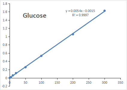
The analytical specifications are calibrated to practical research needs rather than marketing ambitions. The linear calibration range runs from 4 to 300 mg/dL, with a limit of detection of 4 mg/dL. That lower boundary distinguishes basal from pathologically hypoglycemic samples in serum or plasma, while the upper boundary accommodates the hyperglycemic excursions typical of streptozotocin-treated diabetic rodents, glucose-supplemented culture media, and postprandial clinical samples without mandatory dilution. For samples that exceed 300 mg/dL—high-glucose DMEM supplemented with 25 mM glucose, for instance—a straightforward dilution in water brings the concentration back into the linear window. The assay reads at 630 nm, a wavelength accessible on any visible-range microplate reader, including the entry-level models common in teaching laboratories and small biotech startups.
Publication validation is not a footnote for this kit. The product page documents 14 citations in peer-reviewed literature spanning multiple research domains. One publication deployed KTB1300 in a study examining Nanchang University's metabolic research framework, integrating the kit with mitochondrial membrane potential measurements. Other cited work spans glucose quantification in serum, plasma, urine, food, growth media, and plant tissues—a sample compatibility list that exceeds what most glucose assay kits explicitly validate. The fact that 14 independent laboratories, operating under the scrutiny of peer review, chose this specific kit for their glucose measurements provides a level of real-world performance validation that no manufacturer's internal QC data can replicate.
Sample preparation instructions reflect biochemical common sense rather than protocol padding. Fresh samples are recommended, with the option to store material at -80°C for up to one month if immediate assay is not feasible. A brief centrifugation of small vials at low speed before opening prevents cap-associated reagent loss—a small but meaningful detail that suggests the protocol was written by someone who has actually performed the assay on a bench rather than by a technical writer working from a specification sheet. The recommendation to perform several dilutions of the sample to confirm readings fall within the standard value range is standard colorimetric assay discipline, stated explicitly rather than buried in a troubleshooting appendix.
The broader context makes a reliable, interference-resistant glucose assay increasingly essential. Glucose is not merely a clinical diagnostic parameter for diabetes mellitus. It is the primary carbon source for mammalian cell culture, the substrate whose depletion defines the transition from exponential growth to stationary phase, the analyte whose concentration must be held constant in every perfusion and microfluidic system, and the reference point against which glycolytic gene expression changes are interpreted. When a Seahorse analyzer reports extracellular acidification rate, the calculation assumes a known glucose concentration in the assay medium. When a metabolomics core reports glycolytic intermediate pools relative to glucose, the ratio depends on an accurate glucose denominator. When a diabetes researcher measures insulin-stimulated glucose uptake in adipocytes, the assay compares extracellular glucose depletion between stimulated and unstimulated conditions. Each of these workflows inherits whatever systematic error is present in the underlying glucose measurement. A kit that delivers 4 mg/dL detection sensitivity without GOD-POD interference liability, in a two-component format stable at -20°C for 12 months, addresses a need that is simultaneously universal and routinely underestimated.
The 14 citations are not the story. The story is that glucose measurement is the foundation upon which entire experimental edifices are constructed, and a foundation built on an interference-prone enzymatic cascade will eventually produce cracks that reviewers can see. KTB1300 replaces that foundation with a direct chemical condensation that has been validated across serum, plasma, urine, plant tissue, food, and growth medium in laboratories that published their results under peer review. That is not a marketing claim. It is a publication record.
Explore the full specifications, access the protocol, and place your order here: https://www.abbkine.com/product/chekine-micro-glucose-assay-kit-ktb1300/





