


Hic-5 / TGFB1I1: The TGF-β-Inducible Focal Adhesion Hub — And How to Accurately Quantify It with a Sandwich ELISA
There are proteins that sit quietly in the background of pathway diagrams, and then there are proteins like Hic-5 (TGFB1I1) that turn out to be everywhere at once — anchoring focal adhesions to the actin cytoskeleton, shuttling into the nucleus to coactivate steroid receptors, and popping up on every shortlist of mechanically responsive, TGF-β-driven genes. Formally called Transforming Growth Factor Beta-1-Induced Transcript 1 Protein (TGFB1I1), and better known to most cell biologists as Hic-5 (Hydrogen peroxide-inducible clone 5), ARA55 (Androgen Receptor Associated protein of 55 kDa), or TSC-5, this LIM-domain adapter is a multitasking nodal point where extracellular stiffness, growth factor signaling, and nuclear transcription converge. When you need to move beyond mRNA hits and ask "how much Hic-5 protein is actually there, in this sample, reproducibly?" — the Human TGFB1I1 (Hic-5) ELISA Kit — KTE60509 from Abbkine is the tool that takes you from a vague Western band to a hard, interpolated number.
TGFB1I1 / Hic-5: A Molecular Adapter That Refuses to Stay in One Compartment
What makes TGFB1I1 genuinely fascinating — and frustrating to pin down with Western blots alone — is that it plays double-duty in completely different subcellular theaters:
• At focal adhesions and the actin cytoskeleton, Hic-5 uses its four LIM zinc-binding domains to scaffold a growing list of interactors — including ILK, LIMS1/Paxillin, GIT1, TRAF4, PTPN12, CRIP2, SMAD3, and more — positioning it as a key coordinator of mechanotransduction, cell adhesion, migration, and TGF-β / integrin cross-talk.
• In the nucleus, the same protein moonlights as a steroid / nuclear receptor coactivator, enhancing transcriptional activity of the androgen receptor (AR), glucocorticoid receptor (NR3C1), mineralocorticoid receptor, and progesterone receptor — which is exactly why it drew early attention in prostate cancer biology as ARA55.
• At the signaling module level, it feeds back into the Wnt and TGF-β/BMP pathways and may influence monoamine transporter trafficking (SLC6A3, SLC6A4), tying adhesion mechanics to neurotransmitter/neuroendocrine regulation.
Its expression is induced by TGF-β1 (the namesake), mechanical compression/stretch, TNF-α, and H₂O₂ — a signature that places it at the intersection of fibrosis, mechanical injury, airway remodeling (e.g. asthma exacerbations), EMT, and tumor microenvironment stiffening.
Why You Need a Quantitative ELISA for TGFB1I1 (Not Just a Western)
Western blotting is brilliant for showing size, modification shifts, and relative trends. But when the question becomes:
• How much Hic-5 is in this patient serum vs. that one?
• Did my TGF-β1 treatment or stretch protocol produce a 1.8× or 3.2× change across biological replicates?
• Can I correlate Hic-5 levels with a functional readout or clinical score?
— you need absolute quantification, not band-density heuristics. A well-validated sandwich ELISA gives you:
• Specific capture (pre-coated mAb prevents "any LIM protein" confusion)
• Orthogonal confirmation via a second detection epitope (biotinylated antibody)
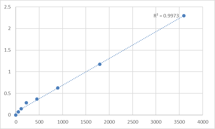
• A standard curve on every plate (so your 450 nm read converts directly into pg/mL or ng/mL)
• Throughput — 96-well format means you can run a cohort, not just a lane-pair
That's exactly what KTE60509 delivers.
Assay Principle: Two-Site Sandwich ELISA, Colorimetric, 450 nm
The Human TGFB1I1 (Hic-5) ELISA Kit (KTE60509) employs the classic, high-specificity sandwich format:
- A mouse anti-human TGFB1I1 capture antibody is pre-coated onto the 96-well microplate.
- Standards and samples (serum / plasma / cell culture supernatant / other biological fluids) are added; any TGFB1I1 present binds.
- After washing, a biotin-conjugated anti-TGFB1I1 detection antibody (recognizing a different epitope) forms the sandwich.
- Streptavidin–HRP binds the biotin.
- TMB substrate produces a blue color ∝ TGFB1I1 amount → stop solution turns it yellow → read Absorbance at 450 nm.
- Interpolate unknowns from the TGFB1I1 standard curve.
The entire assay runs ~3–5 hours door-to-door (depending on user experience), making it realistic to pair with a treatment time-course or a patient-cohort plate in a single day.
Kit Contents & Practical Specs at a Glance
Feature Detail
Target Human TGFB1I1 / Hic-5 / ARA55 (UniProt: O43294, Gene ID: 7041)
Format 96-well sandwich ELISA, pre-coated capture antibody
Detection Biotinylated detection Ab → Streptavidin-HRP → TMB, 450 nm read
Sample Types Serum, Plasma, Cell culture supernatants, Other biological fluids
Assay Duration ~3–5 hours (multi-step standard protocol)
Specificity High sensitivity for human TGFB1I1; no significant cross-reactivity with analogues reported
Status For Research Use Only — NOT for human/clinical diagnosis
Where Quantifying TGFB1I1 / Hic-5 Actually Moves the Needle
- Fibrosis, TGF-β biology & mechanotransduction
Hic-5 is a direct TGF-β1-responsive gene and a focal adhesion mechanosensor proxy. Quantifying it in cell supernatants / lysates / (potentially) surrogate fluids gives you a protein-level correlate to complement p-SMAD2/3 and stiffness assays — especially in pulmonary, renal, or hepatic fibrotic models. - Prostate & androgen-driven cancer contexts
As ARA55, Hic-5 modulates AR transcriptional output and stromal–epithelial crosstalk. An ELISA lets you screen culture conditioned media or tissue extracts across lines, knockdowns, and drug treatments without burning gels. - Airway remodeling & asthma exacerbations
RNA-seq + qPCR work has already tied TGFB1I1 mRNA to mechanical compression and rhTGF-β1 in human bronchial epithelial cells — and to bronchoconstriction in patient contexts. A protein-level readout kit lets you ask whether Hic-5 levels track with severity or response to mechanomodulatory drugs. - EMT, migration & invasion assays
Because Hic-5 localizes to focal adhesions and regulates actin stress-fiber dynamics and adhesion turnover, its abundance often shifts during EMT transitions — but those shifts need to be quantified across replicates, not guessed from one blot. - iPSC / stromal differentiation models
Tracking TGFB1I1 as a differentiation/stromal activation marker across timepoints becomes trivial when every timepoint is a 50 µL sample into a 96-well plate.
A Few "Pro-Tips" That Keep Your TGFB1I1 ELISA Clean
• Never mix lots — keep standards, detection Ab, and HRP from the same box.
• Warm everything to RT (≥30 min) before you open tubes — cold reagents + condensation = micropipette inaccuracy.
• Include a serially diluted standard curve on every plate — never rely on "it looks like yesterday."
• If you're coming from tissue homogenates, clarify well (centrifuge) and consider a small pilot dilution series to make sure your matrix doesn't push the signal outside the linear range.
• Remember: Research Use Only — this is discovery data, not a diagnostic test.
The Bottom Line
TGFB1I1 / Hic-5 is one of those proteins that keeps showing up in the places that matter most — where mechanics, growth factors, and transcription collide. But naming it in a pathway diagram isn't the same as measuring it with confidence. The Human TGFB1I1 (Hic-5) ELISA Kit — KTE60509 from Abbkine gives you a pre-coated sandwich ELISA platform that's specific, reproducible, and fast enough (~3–5 h) to scale across conditions, cohorts, and replicates — so your next Hic-5 figure is built on numbers, not a "dark band vs. light band" eyeball estimate.
Product Reference: KTE60509 – Human Transforming growth factor beta-1-induced transcript 1 protein (TGFB1I1) ELISA Kit
Learn more and order: https://www.abbkine.com/product/human-transforming-growth-factor-beta-1-induced-transcript-1-protein-tgfb1i1-elisa-kit-kte60509/


