


From Testis to Retina: The Dual-Identity Protein SPATA7 and Why You Need a Dedicated Sandwich ELISA to Quantify It
Some proteins refuse to stay in their lane—and SPATA7 (Spermatogenesis-Associated Protein 7) is the ultimate example. First cloned from testis cDNA libraries and named for a reproductive process, it turned out to be highly expressed in the brain and retina, localizes to the photoreceptor connecting cilium, and—when mutated—ranks among the verified causative genes for Leber Congenital Amaurosis (LCA3) and autosomal recessive juvenile retinitis pigmentosa. In other words, the same gene flagged in spermatogenesis literature now sits on the list of retinal ciliopathy targets that ophthalmologists and geneticists screen in severely visually impaired children. That dual identity—testis and ciliary sensory neuron—is exactly why SPATA7 demands rigorous, protein-level measurement rather than a hand-waved band on a gel. The Human Spermatogenesis-associated protein 7 (SPATA7) ELISA Kit (KTE60543) from Abbkine gives labs a two-site sandwich ELISA that converts this elusive, coiled-coil-rich ciliary protein into a hard, interpolated number you can actually use for statistics, cohort comparison, and mechanism work.
SPATA7: A "Spermatogenesis" Name That Hides a Ciliary Emergency
Despite the name, SPATA7 (UniProt: Q9P0W8; aliases: HSD-3.1 / HSD3 / LCA3) is conserved from sea urchin to human and is most abundant in testis but also strongly expressed in retina, brain, and RPE. Sequence analysis reveals no classic catalytic domain—its functional anchor is a predicted N-terminal coiled-coil domain (aa 81–109) that mediates microtubule association and ciliary localization.
The modern model of SPATA7 function centers on the connecting cilium (CC) of the photoreceptor:
• SPATA7 localizes to the CC/transition zone area and decorates the ciliary axoneme.
• It directly interacts with RPGRIP1 via that coiled-coil region; in Spata7 loss-of-function, RPGRIP1 mislocalizes from the CC into the inner segment.
• Rhodopsin (RHO) trafficking across the IS→OS border breaks down, leading to apoptotic photoreceptor death and progressive degeneration.
Clinically, biallelic SPATA7 mutations account for an estimated ~1.7–4.6% of LCA patients (population-dependent) and present as either severe congenital/early infantile LCA or a juvenile RP/EOSRD phenotype—roving nystagmus, extinguished ERGs, severely constricted visual fields, and progressive outer retinal atrophy.
Why "Just Western" Isn't Enough for SPATA7
SPATA7 is non-abundant in the sense that it's a structural/trafficking adaptor, not a housekeeping enzyme churned out by the ton. That means:
• A Western blot often teeters between "visible" and "too faint for densitometry."
• Loading normalization (Actin/Tubulin) can dominate the error budget if total protein is slightly off.
• You can't easily compare multiple cohorts, iPSC-derived retinal organoid batches, or serum/plasma surrogates with a single gel-image.
A well-validated sandwich ELISA bypasses those bottlenecks by giving you:
• Two independent epitopes (capture Ab + biotin detection Ab) → higher specificity, lower ambiguity
• A plate-based standard curve run with every assay → corrects plate-to-plate drift automatically
• Objective OD → concentration readout at 450 nm (TMB/HRP system) instead of "darker band vs. lighter band"
Assay Principle: The KTE60543 Sandwich ELISA in a Nutshell
The kit employs a standard quantitative sandwich format:
- A microplate is pre-coated with a capture antibody specific for human SPATA7.
- Standards + samples (serum / plasma / tissue homogenates / cell culture supernatants / other biological fluids) are added; SPATA7 binds.
- After washing, a biotinylated anti-SPATA7 detection antibody binds a different epitope → Streptavidin–HRP attaches.
- TMB generates color ∝ bound SPATA7; stop solution terminates the reaction (yellow readout).
- Absorbance at 450 nm → interpolate unknowns from the SPATA7 standard curve.
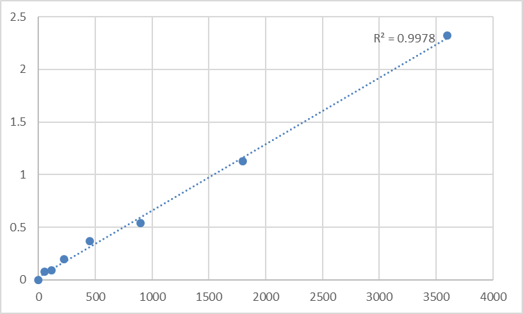
Reported performance benchmarks you'll commonly see cited for kits in this family include:
• Detection range: roughly 0.156–10 ng/mL class (plate-dependent)
• Limit of detection: on the order of ~0.094–0.156 ng/mL
• Intra-assay CV: often ≤ 6.5%; Inter-assay CV: ≤ 8.9%
• Recovery: close to ~96–102% in validated matrices
• Assay duration: ~3–5 hours (depends on user/step timing)
(Always confirm the exact range/CV for your lot/manual; these are representative validated-class numbers distilled from supplier datasheets.)
Where Quantifying SPATA7 Actually Moves the Needle
- Retinal ciliopathy & photoreceptor trafficking studies
Track SPATA7 protein levels in iPSC-derived retinal organoids, 3D photoreceptor cultures, or RPE-photoreceptor co-cultures under CRISPR knockout/rescue, AAV-Spata7 gene therapy conditions, or chemical stress (oxidative / proteostatic insults). Protein-level readouts make rescue quantitation defensible.
- Spermatogenesis & male germ cell stress
Because SPATA7 originated as a testis-associated factor, it remains relevant in germ-cell biology: spermatogenic stage-specific expression, heat stress, toxin exposure, or hormonal manipulation models. An ELISA lets you run multiple germ-cell fractions or tubule lysates in one plate without burning gels.
- Surrogate fluid / biomarker feasibility
Although SPATA7 is chiefly a tissue/cell protein, any effort to explore circulating exosomal or shed photoreceptor markers eventually needs a specific, low-cross-reactivity capture assay—which is exactly what a pre-validated sandwich ELISA framework provides as a foundation.
- Genetic validation pipelines
If you're engineering SPATA7 edits (CRISPR frameshift, splice rescue, AAV-Biallelic conversion), show the world more than "absent band": report % SPATA7 protein remaining ± SEM derived from a calibrated curve.
A Quick Lab-Checklist to Protect Your Data
• Clarify tissue/cell lysates well before loading into the plate (centrifuge; keep cold).
• Bring all reagents to RT before opening; avoid condensation on tips/tube walls.
• Always run the full standard curve on every plate—never reuse a curve from a previous run.
• Include a blank-matrix control (your lysis/RPMI/base medium without cells) so background subtraction is honest.
• Store per manual (typically 4°C / -20°C as instructed); protect from frost-free freezer cycles; avoid unnecessary freeze–thaws by aliquoting if your workflow is high-frequency.
The Bottom Line
SPATA7 started its career in the literature as a "spermatogenesis-associated" clone and grew into a bona fide LCA3 / retinal ciliopathy gene that bridges microtubule-associated transport, connecting-cilium integrity, and RPGRIP1 trafficking. Measuring it properly means respecting that biology—and that means moving from qualitative blots to a calibrated, sandwich ELISA workflow. The Human Spermatogenesis-associated protein 7 (SPATA7) ELISA Kit — KTE60543 from Abbkine is built for exactly that job: pre-coated capture, biotin detection, HRP-TMB readout at 450 nm, and a full buffer/reagent system so your numbers hold up across replicates, conditions, and users.
Product Reference: KTE60543 – Human Spermatogenesis-associated protein 7 (SPATA7) ELISA Kit
Learn more and order: https://www.abbkine.com/product/human-spermatogenesis-associated-protein-7-spata7-elisa-kit-kte60543/
(For Research Use Only; not for diagnostic or therapeutic use in humans.)


