


Beyond the "Repulsion" Myth: Why Human SLIT3 Is a Pro-Angiogenic Secreted Cue You Need to Quantify — And How the KTE60580 Sandwich ELISA Makes That Possible
Few molecules in modern cell biology carry a name that is more misleading than Slit3. The "Slit" family was baptized in the 1990s as a set of large, secreted repulsive axon-guidance proteins that told growing neurons "don't cross here." But the deeper we've dug, the clearer it became that SLIT3 (Slit homolog 3, UniProt: O75094, Gene ID: 6586) is not just a neuronal repellent — it is a potent, secreted glycoprotein (≈ 170–200 kDa) that gets exported into the extracellular space, decorates the matrix, and talks to endothelial cells and mural-cell precursors through ROBO receptors (especially ROBO4) to coordinate vascular network formation, lymphatic development, diaphragm/kidney organogenesis, and even engineered-tissue vascularization. In short: SLIT3 is one of those rare proteins whose biology refuses to stay in its historical lane, and that makes having a reliable, protein-level readout absolutely essential. The Human Slit homolog 3 protein (SLIT3) ELISA Kit (KTE60580) from Abbkine is purpose-built for exactly that job — a sandwich ELISA that lets you move from "I think my supernatant is richer in SLIT3" to a calibrated concentration (ng/mL) you can plot, normalize, and defend.
SLIT3: From "Don't Touch" Signal to Paracrine Pro-Vascular Factor
Structurally, SLIT3 is built around leucine-rich repeats (LRRs), EGF-like repeats, and a C-terminal domain — classic modular scaffolds for protein–protein interaction at the cell surface and in the ECM. Developmentally, Slit3 knockout mice don't collapse the CNS in the way Slit1/2 deletions do; instead, they present with aortic arch anomalies, lymphatic defects, and failures in diaphragm and kidney morphogenesis, proving SLIT3's action radius is heavily weighted toward non-neuronal developmental plumbing.
The real pivot came when vascular biologists showed that SLIT3 is expressed and secreted by endothelial cells and vascular smooth muscle cells, and that it stimulates endothelial proliferation, motility, chemotaxis, and capillary-network formation in vitro — with a potency that can rival VEGF in specific bioassay contexts. Its cognate receptor ROBO4 is a key endothelial sensor: SLIT3–ROBO4 signaling stabilizes/guides neovessel assembly, and in 3D tissue-engineering models, knocking down SLIT3 in pericyte-like MSCs disrupts organized EC networks, while knocking down ROBO4 in ECs abolishes functional human vessel formation in vivo.
The takeaway for your bench: if you work on angiogenesis, engineered vascularized grafts, MSC paracrine screens, lymphatic development, or congenital cardiopulmonary defects, pretending SLIT3 is "just an axon thing" will blind you to a secretory signal that may be driving your phenotype.
Why a Sandwich ELISA — and Why KTE60580
Western blots are still great for confirming size or checking gross expression trends, but SLIT3 is a secreted, glycosylated, ECM-associated protein whose biologically relevant readout is often how much soluble (or mildly matrix-bound) SLIT3 is accumulating in media, serum, plasma, or tissue extracts — not just whether a band exists in a whole-cell lysate. A two-site sandwich ELISA wins here because:
• Two independent epitopes (pre-coated capture Ab + biotin detection Ab) give you far higher specificity than a single "one-Ab" colorimetric dip.
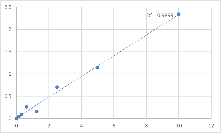
• A recombinant-standard curve on every plate converts OD₄₅₀ into absolute ng/mL, so "day 14 vs. day 0" or "clone A vs. clone B" becomes a statistics problem, not an artistic-interpretation problem.
• The format is intrinsically high-throughput friendly: 48T/96T layouts let you run time courses, dose responses, or patient-cohort batches without chaining yourself to a gel rig.
The KTE60580 kit implements exactly this architecture:
- Microplate pre-coated with anti-human SLIT3 capture antibody.
- Standards / samples added → SLIT3 captured.
- Biotinylated anti-SLIT3 detection antibody → Streptavidin–HRP → TMB (blue) → stop solution (yellow).
- Read at 450 nm; color intensity ∝ [SLIT3]; interpolate from the standard curve.
Performance Snapshot (the Numbers You'll Actually Cite)
Across distributor/technical summaries that mirror the kit's design space, you'll commonly see:
• Detection range: 0.156 – 10 ng/mL
• Limit of Detection (LOD): in the ballpark of ~0.06 – 0.08 ng/mL
• Intra-assay CV: typically < 10% (often quoted ≤ ~6–8%)
• Inter-assay CV: typically < 12%
• Spike-recovery: e.g., serum ~93–102%, EDTA plasma ~98–105%, heparin plasma ~86–93% (expect matrix-specific offsets; run your own controls)
• Samples: serum, plasma (EDTA/heparin), tissue homogenates, cell culture supernatants, cell lysates, other biological fluids (follow the manual's prep/clarification advice)
These are the "materials & methods" numbers that tell reviewers: this isn't a band-count — it's a calibrated assay.
Where Quantifying SLIT3 Actually Moves Your Story Forward
- MSC / pericyte paracrine screens & engineered-tissue vascularization
If you're testing whether a mesenchymal stem cell / pericyte-like population supports EC network assembly, SLIT3 is one of the few secreted guidance cues worth putting on a spreadsheet. KTE60580 lets you correlate ng/mL SLIT3 in conditioned media with network length, lumen formation, or in vivo graft perfusion — turning a "maybe secretory factor" into a quantified contributor.
- Developmental / congenital vascular-defect models
Congenital anomalies linked to aortic arch/lymphatic/diaphragm phenotypes (where Slit3 KO mice live) benefit from a circulating or tissue-level SLIT3 readout when you're comparing mutants vs. littermates or testing rescue constructs.
- Tumor microenvironment & lymphangiogenesis
SLIT–ROBO signaling is context-dependent in cancer (can suppress invasion via repulsion in some settings, can support vascular/lymphatic patterning in others). If your tumor model secretes SLIT3, measure it — protein concentration beats mRNA fold-change when the molecule lives outside the cell.
- Drug / siRNA / AAV screens on endothelial or smooth-muscle secretomes
Running a conditioned-medium campaign? ELISA is often the fastest way to close the loop: did the manipulation actually shift what gets secreted, or did it just change intracellular pools?
A Minimal "Clean-Data" Workflow You Can Copy-Paste into Your Notebook
- Thaw reagents to RT (≥ 30 min) before opening; keep TMB protected from light.
- Clarify supernatants/media (centrifuge, no cell debris) and, for tissue, do a proper homogenization + centrifuge + dilute to stay inside the 0.156–10 ng/mL working range.
- Add standards + samples per protocol; incubate; wash per recommended cycle (don't shortcut the soak/wash volume).
- Develop with TMB; stop uniformly (same order, same timing); read 450 nm promptly.
- Fit a 4-PL (four-parameter logistic) if your software allows; export ng/mL; back-calculate dilution factors; log the plate map.
Bottom Line
SLIT3 is the molecule that outgrew its own name: born as a "repulsive axon cue," it turned out to be a secreted pro-angiogenic paracrine factor that helps decide whether new vessels form, how lymphatic plumbing develops, and whether engineered tissues survive after implant. If your project touches any of those questions, you owe it to yourself to measure SLIT3 like a scientist — with a calibrated standard curve, not a gel-shadow. The Human Slit homolog 3 protein (SLIT3) ELISA Kit — KTE60580 from Abbkine is the tool that gets you from "there might be SLIT3 here" to "there are X ng/mL, and here's the curve that proves it."
Product Reference: KTE60580 – Human Slit homolog 3 protein (SLIT3) ELISA Kit
Learn more and order: https://www.abbkine.com/product/human-slit-homolog-3-protein-slit3-elisa-kit-kte60580/
(Research Use Only — not for diagnostic procedures in humans.)


