




The Midline Repellent Goes Soluble: Why Human SLIT1 Demands a Protein-Level Readout — And How the KTE60582 Sandwich ELISA Delivers
The word "Slit" still conjures one very specific image for developmental neurobiologists: a growing axon approaching the ventral midline of the neural tube, encountering a cloud of secreted repellent, and veering sharply away. That foundational midline-screen discovery in Drosophila — where slit mutations caused commissural axons to keep crossing instead of stopping — is textbook material. But the vertebrate story is far more interesting than a simple "no trespassing" sign. Humans carry three Slit homologs (SLIT1, SLIT2, SLIT3), and SLIT1 (Slit homolog 1, UniProt: O75041, aliases: MEGF4 / SLIL1 / KIAA0813 / Slit-1) is the family's most brain-restricted, secreted extracellular matrix-associated member — a bulky (~1534-aa, ~190 kDa precursor) leucine-rich repeat protein that diffuses from producing cells, decorates the ECM via heparan sulfate proteoglycans (e.g., glypican-1), and talks to ROBO receptors to steer migrating neurons, repel olfactory axons, and sculpt forebrain wiring. Because it is actively secreted rather than trapped inside a cell, the biologically relevant question isn't just "is SLIT1 there?" — it's how much soluble SLIT1 is your culture supernatant, CSF, serum, or tissue extract actually producing? The Human Slit homolog 1 protein (SLIT1) ELISA Kit (KTE60582) from Abbkine is built to answer exactly that, with a two-site sandwich ELISA that turns this iconic guidance cue into a calibrated number you can plot, normalize, and put in a table with error bars.
SLIT1's Real Job: A Secreted Chemoarchitectural Cue, Not a Static Marker
Structurally, SLIT1 is built around four N-terminal leucine-rich repeat (LRR) domains, an EGF-like repeat array (the classical Slit cysteine-knot/CT domain region), and a C-terminal cystine-knot (CT) domain — a modular architecture designed for long-range extracellular diffusion, HSPG binding, and multivalent protein–protein interaction. Its canonical receptors are Roundabout (ROBO1/ROBO2); the Slit → ROBO engagement at the growth cone triggers local actin reorganization that collapses or repels advancing processes. In the mammalian forebrain, SLIT1 (together with SLIT2) helps enforce midline exclusion — e.g., repelling and preventing recrossing of olfactory bulb–projecting axons — and contributes to interneuron migration and cortical circuit assembly. Northern and expression surveys consistently flag SLIT1 as predominantly expressed in adult forebrain, with strong fetal brain signal and detectable levels in lung and kidney, but far more restricted than the broadly expressed SLIT2 or the endothelial-weighted SLIT3.
The translational kicker is this: because SLIT1 is secreted and ECM-associated, its functional dose matters in contexts that go well beyond a single immunostaining section — conditioned-media screens, organoid-conditioned harvests, surgical aspirates or CSF, even attempts at surrogate circulatory biomarkers for neurodevelopmental or neuro-oncology conditions. And that is where a gel band fails you.
Why "Band Intensity" Breaks Down — And Why Sandwich ELISA Wins
A Western blot is fantastic for showing size, cleavage state, and spatial validation ("yes, the ~190 kDa band is in the forebrain lane and gone in the KO"). But when the claim becomes quantitative —
"Condition A drives a ~2.5-fold increase in secreted SLIT1 over 48 h relative to control…"
— a densitometric band on a membrane can't carry that weight alone. A sandwich ELISA solves three structural problems at once:
- Two independent epitopes (pre-coated capture Ab + biotin detection Ab) give much higher specificity than any single antibody approach — critical for a large, repetitive LRR protein where false cross-reactivity is a real danger.
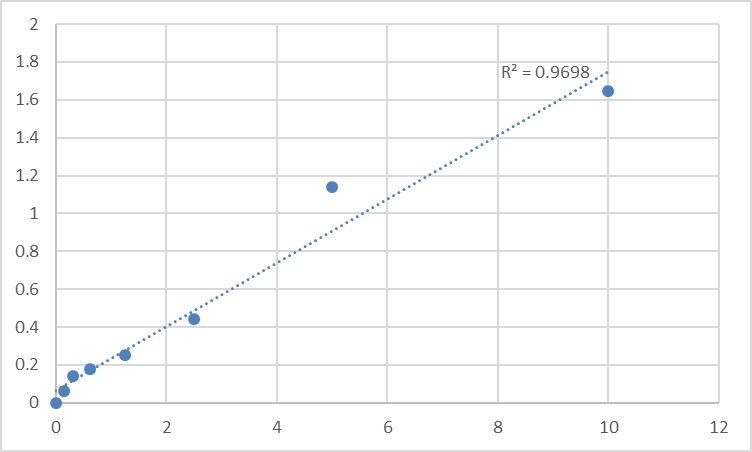
- A recombinant standard on every plate converts absorbance into absolute concentration (ng/mL), killing the "was my exposure linear?" anxiety.
- Throughput — 48T/96T formats mean you can run a time-course × dose × replicate matrix in one sitting instead of burning an entire week on gels.
Assay Principle: The KTE60582 Sandwich ELISA, Step by Step
The Human SLIT1 ELISA Kit (KTE60582) uses the classic, field-standard two-site format:
- A microplate is pre-coated with a capture antibody specific for human SLIT1.
- Standards and samples (serum / plasma / cell culture supernatants / tissue homogenates / other biological fluids) are added; any SLIT1 present binds.
- After washing, a biotin-conjugated anti-SLIT1 detection antibody (recognizing a different epitope) forms the sandwich complex.
- Streptavidin–HRP binds the biotin; TMB substrate produces blue color ∝ bound SLIT1.
- Stop solution turns it yellow; read Absorbance at 450 nm; interpolate unknowns from the SLIT1 standard curve.
The assay runs ~3–5 hours door-to-door (depends on user experience and incubation timing) and is strictly for research use — not for clinical diagnosis.
Performance Snapshot (the numbers you'll cite)
Across supplier summaries that mirror this kit's design space, you'll typically see:
Parameter Typical Specification
Target Human SLIT1 / Slit-1 (UniProt: O75041)
Format 96-well (also 48T) sandwich ELISA, pre-coated capture
Detection Biotin-Ab → Streptavidin–HRP → TMB, 450 nm read
Dynamic Range 0.156 – 10 ng/mL (standard 7-point series)
Sensitivity / LOD ~0.06–0.08 ng/mL
Intra-Assay CV < 10% (often ≤ ~5%)
Inter-Assay CV < 12% (often ≤ ~7%)
Recovery (serum/plasma) Serum ~93%, EDTA plasma ~87%, Heparin plasma ~94% (verify in your own matrix)
Samples Serum, plasma, cell culture supernatants, tissue homogenates, other biological fluids
Assay time ~3–5 hours
(Always confirm exact range/CV for your lot and protocol in the official Abbkine manual.)
Where Quantifying SLIT1 Actually Advances the Story
- Neural development & migration assays
If you're tracking neuron/neuronal-precursor migration (olfactory neurogenesis, interneuron displacement, forebrain explant models), secreted SLIT1 in the medium or matrix extract is the relevant readout — not just an intracellular tag. The ELISA lets you correlate ng/mL SLIT1 output with migratory speed, trajectory stats, or manipulative treatments (ROBO-Fc, siRNA/sgRNA, small molecules).
- Slit–ROBO cancer & invasion studies
Although SLIT2 dominates the tumor-biology literature, SLIT1 shows up in certain cancer cell secretomes and CNS-tumor microenvironments, where Slit–ROBO signaling can either suppress or enable invasion depending on receptor context and cellular origin. Quantifying secreted SLIT1 avoids the trap of equating "intracellular mRNA" with "what the tumor is broadcasting."
- 3D brain organoids & engineered neural tissue
Organoid-conditioned media are noisy and low-volume. A sandwich ELISA is often the only practical way to get a longitudinal, multi-timepoint handle on how differentiation protocols, mechanical strain, or CRISPR edits affect guidance-cue secretion without sacrificing the organoid.
- Neuroinflammation / innate immune migration
Yes — ROBO–Slit signaling also modulates leukocyte chemotaxis and macrophage migration in select contexts. If your model implicates SLIT1 in "don't come here" signaling at a vascular or meningeal interface, the protein concentration in supernatant is the variable you need locked down.
- sgRNA/AAV validation pipelines
Editing SLIT1 (or ROBO1/2)? Don't just show "band lighter." Report % SLIT1 protein remaining ± SEM derived from a calibrated curve — reviewers notice the difference.
Quick Best-Practice Checklist
• Clarify before you load: spin supernatants / homogenates to remove debris (cloudy samples = scattered A₄₅₂ = fake highs).
• Warm everything to RT ≥ 30 min before opening; keep TMB dark.
• Run the full standard curve on every plate — never reuse a curve from a different day/lot.
• If your sample reads above range, dilute in the recommended buffer, not water, and back-calculate with the dilution factor.
• Store unopened at 2–8°C; protect unused wells in the foil bag with desiccant at 4°C once opened.
The Bottom Line
SLIT1 earned its reputation as the Drosophila midline's "keep out" signal, but in human biology it's a secreted, HSPG-bound architect that shapes forebrain wiring, neuronal migration, and the extracellular logic of an entire tissue. If your project touches that logic, you owe it to yourself to measure SLIT1 where it actually lives — outside the cell, in solution and matrix — with a calibrated, two-antibody sandwich assay instead of a guess on a membrane. The Human Slit homolog 1 protein (SLIT1) ELISA Kit — KTE60582 from Abbkine is the tool that gets you there: pre-coated capture, biotin detection, HRP–TMB readout at 450 nm, and a 0.156–10 ng/mL window that's sensitive enough for real biofluid and supernatant work.
Product Reference: KTE60582 – Human Slit homolog 1 protein (SLIT1) ELISA Kit
Learn more and order: https://www.abbkine.com/product/human-slit-homolog-1-protein-slit1-elisa-kit-kte60582/
(For Research Use Only; not for diagnostic use in humans.)





