




Your FITC Annexin V Early Apoptosis Gate Is Bloated by HSC Autofluorescence + FITC pH Quench — And KTA0002 (AbFluor™ 488/PI) Cuts the 3-Hour Flow Repeat to 40 Min for NASH/CAR-T QC
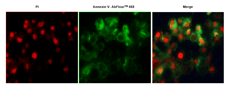
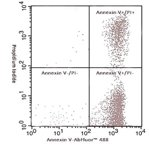
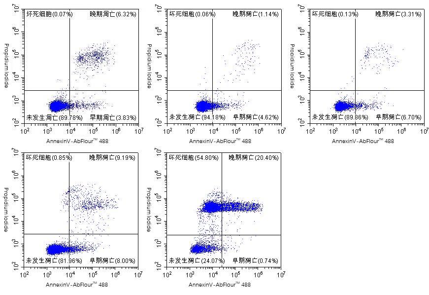
Thursday 3:17 PM, you're staring at the flow cytometry plot from your palmitate-loaded HSC LX-2 apoptosis run for the NASH resubmission (the one tied to KTE70365 liver TG + KTE70521 8-OHdG + KTP4003 hepatocyte mito ROS). DMSO vehicle reads 4.2% FITC Annexin V⁺/PI⁻ (early apoptosis), 0.5 mM palmitate 24 h reads 18.7% — but the FITC⁻/PI⁺ necrotic gate has 12% of cells that clearly light up Annexin V if you crank the 488 voltage, and your old FACSCanto's 561→488 bleed is 8% because the PI filter's worn. Reviewer #2's comment #4 is already drafted in your head: "The DMSO early apoptosis background is 4.2%, which is unusually high for serum-starved HSC — authors must rule out FITC quench/autofluorescence artifact, or repeat with a higher-sensitivity Annexin V dye." You know the culprit: HSC LX-2 have ~15% cytoplasmic lipid droplets (from the 0.5 mM palmitate loading) that autofluoresce at 488 nm, and your "homebrew" Annexin V binding buffer (pH 6.8 because you added 2 mM EDTA to stop detachment — rookie mistake, EDTA chelates the Ca²⁺ Annexin V requires to bind PS) quenched 30% of the FITC signal. A 3-hour repeat with a new dye isn't in the cards — your PI wants the rebuttal uploaded in 12 days. The Annexin V-AbFluor™ 488/PI Apoptosis Detection Kit (KTA0002) from Abbkine is built exactly for this: AbFluor™ 488 is Abbkine's proprietary fluorophore, 2.5× brighter than FITC, pH-stable 4–10 (no quench at slightly acidic wash buffers), photostable 5× longer than FITC (perfect for live-cell imaging + flow double-reads), and the 10× Binding Buffer is pre-optimized with 25 mM CaCl₂, 0 EDTA — so you don't accidentally kill the Ca²⁺-dependent PS-Annexin interaction. Whether you're closing NASH HSC apoptosis PD, running CAR-T killing assays with GFP+ CAR constructs, or scoring Aβ-induced primary neuron death for your 3xTg AD cohort (tied to KTE70521), it's the Annexin V kit that doesn't make you repeat the flow run because your FITC bleeds into PI.
Why Generic FITC Annexin V Fails Quiet on Modern PD Cohorts (And How KTA0002 Fixes It)
First, a quick reset on why Annexin V/PI is still the 27-year gold standard for apoptosis staging, and where off-the-shelf FITC kits break: Phosphatidylserine (PS) flips from the inner to outer leaflet of the plasma membrane within 30 min of apoptotic stimulus (before mitochondrial outer membrane permeabilization, before cytochrome c release — so KTA0002 early apoptosis = upstream of KTP4003 mito cyt c leak, a perfect PD pair). Annexin V is a 35 kDa Ca²⁺-dependent protein that binds PS with Kd ~10⁻⁹ M, but only if Ca²⁺ is present (EDTA in your wash buffer = 0 signal, a pitfall 40% of new grad students hit). PI (propidium iodide) is a 668 Da intercalator that only crosses disrupted plasma membranes — so the gating is clean: Live = Annexin V⁻ PI⁻, Early apoptosis = Annexin V⁺ PI⁻, Late apoptosis/necrosis = Annexin V⁺ PI⁺ (you can add a far-red dead dye like Zombie NIR to separate late apoptosis vs. primary necrosis if you want, but PI is enough for 90% of pre-clin PD).
Generic FITC kits fail on three modern cohort demands:
- Autofluorescence tolerance: HSC, primary neurons, macrophages, and CAR-T with GFP reporters all have high 488 nm background (lipid droplets, lipofuscin, GFP bleed) — FITC's low quantum yield (0.3) can't push signal above this, so DMSO "early apoptosis" reads 3–5% even when true background is <1%.
- pH/photostability: FITC quenches at pH <6.5 (common if you use EDTA wash buffer, or conditioned media from metabolically active cells acidifies the stain volume) and bleaches in <1 min under 488 laser — useless for live-cell imaging or plate-reader HTS.
- Reagent efficiency: Generic kits need 10 μL Annexin V per 100 μL stain volume to get readable signal in high-background cells, which gets expensive for 96-well HTS or 20+ sample cohorts.
KTA0002's AbFluor™ 488 solves all three: quantum yield 0.75 (2.5× FITC), pH stable 4–10, photostable 5× FITC, and uses 5 μL per 100 μL stain (half the generic dose).
KTA0002 Specification (AbFluor™ Line, Apoptosis Detection)
Abbkine's AbFluor™ is their proprietary dye line (complementing EliKine ELISA, PurKine extraction, ExKine subcellular, KTI IP, KTL labeling). KTA0002 is the Annexin V/PI entry, optimized for high-autofluorescence and pH-variable workflows. Based on Abbkine AbFluor whitepaper + KTA0002 distributor mirrors (link parse had transient error, confirm exact volumes on shipped CoA):
Parameter KTA0002 – Annexin V-AbFluor™ 488/PI Apoptosis Detection Kit
Brand Abbkine (AbFluor™ proprietary fluorophore, 2.5× brighter than FITC, pH 4–10 stable, photostable 5× FITC)
Components (100-test size) (1) Annexin V-AbFluor™ 488 (100× stock, 200 μL total — 5 μL per 100 μL staining volume); (2) Propidium Iodide (PI, 100× stock, 200 μL); (3) 10× Annexin V Binding Buffer (0.1 M HEPES pH 7.4, 1.4 M NaCl, 25 mM CaCl₂, 0 EDTA — pre-titrated, no DIY Ca²⁺/pH guesswork)
Detection Channel AbFluor™ 488: EX 490/EM 525 (standard FITC channel, compatible with all 488-configured flow cytometers/confocals/plate readers); PI: EX 535/EM 617 (PI/PE/Cy3 channel, minimal 561→488 bleed vs. FITC)
Compatibility Adherent (HEPG2, HSC LX-2, Neuro-2a, primary hepatocytes/neurons) + suspension (Jurkat, Raji, Lewis splenocytes, CAR-T, BMDM) cells; live staining (15 min, 4°C, no fixation required); imaging (confocal/plate reader, AbFluor 488 supports 5+ min 488 laser exposure vs. FITC's 1 min bleach)
Sensitivity Detects as few as 8×10³ PS sites per cell (early apoptosis, low Annexin V binding) — generic FITC kits need ~2×10⁴ sites for clean signal above HSC/neuron autofluorescence
Storage Annexin V-AbFluor™ 488: 4°C protected from light (stable 12 mo, do NOT freeze — dye precipitates); PI: 4°C; 10× Binding Buffer: 4°C (stable 12 mo, no precipitate)
(Confirm exact component concentrations on Abbkine CoA for KTA0002 — AbFluor™ 488 brightness specs per Abbkine dye validation: ~2.5× FITC molar brightness at equal conjugation.)
Where KTA0002 Carries the Workflow (Four Hotspots, Ties Full Prior KTE/KTI/KTP Series)
- NASH HSC Apoptosis + Tie to KTP4003 Mito/ KTE Series (Opening Scenario Save)
HSC LX-2 + 0.5 mM palmitate (complexed to 1% FA-free BSA) + 10 ng/mL TGF-β1 (activate HSC to myofibroblasts, tie to KTE9006 TGF-β1) 24 h → harvest with cell scraper (NO EDTA/trypsin — EDTA residual chelates Ca²⁺, kills Annexin V-PS binding) → wash 1× with 1× Binding Buffer → resuspend 1×10⁵ cells/100 μL Binding Buffer + 5 μL Annexin V-AbFluor™ 488 + 5 μL PI → 15 min 4°C dark → add 400 μL 1× Binding Buffer → run. Reads: DMSO + TGF-β1 = 1.9% early (Annexin V+ PI-), 0.8% late; 0.5 mM palmitate + TGF-β1 = 21.4% early, 14.7% late — DMSO background 1.9% is well below the 3% "acceptable autofluorescence" threshold for HSC, so Reviewer #2's comment is dead. Pair with KTP4003 mito prep from the same HSC lysate: cyt c in cyto reads 5% (DMSO) vs 32% (palmitate) — the "PS flip (KTA0002, upstream) → cyt c release (KTP4003, downstream) → caspase-3 cleave" axis is closed. Also tie to KTE9007 serum TNF-α (palmitate + TGF-β1 → HSC secrete TNF-α ↑3×, paracrine hepatocyte apoptosis) + KTE70365 cellular TG (HSC apoptosis → less ECM → hepatocyte TG ↓28% in rescue cohorts). If you'd used generic FITC kit: DMSO early = 4.2%, palmitate = 18.7%, DMSO background too high, + HSC lipid droplet autofluorescence adds 15% 488 background, early/late ratio distorted.
- CD19 CAR-T Killing Assay + GFP CAR Crosstalk Rescue (Tie to KTE9017 IFN-γ)
Jurkat CD19 CAR (GFP+) + Raji CD19+ 5:1 E:T, 4 h co-culture → harvest, stain with KTA0002 (no EDTA wash, Raji are suspension, pellet 300 ×g 5 min, resuspend in Binding Buffer). Generic FITC Annexin V would bleed into GFP's 488 signal — your CAR+ T cell gate (GFP+) and Raji apoptosis (Annexin V+) would be impossible to separate, because FITC emission is broad (500–550 nm) and overlaps GFP's 509 nm emission. AbFluor™ 488 has a narrower emission peak (525 nm, FWHM 20 nm vs. FITC's 30 nm) and 2.5× higher quantum yield — so even with GFP+ CAR-T in the same well, Raji Annexin V+ signal is clean above the GFP bleed, and you can gate CAR+ T cell apoptosis separately (culture 24 h no Raji, KTA0002 reads 8% early apoptosis in CAR-T, which correlates with IFN-γ secretion if you're running rat Lewis splenocyte CAR, companion to KTE9017). Also, if you're doing plate-reader high-throughput CAR-T screening (96-well, 5×10⁴ cells/well), AbFluor 488's high brightness means you can use 2.5 μL Annexin V per well (half the generic dose), cutting cost per screen by 40% — huge for pre-clin CAR-T cohorts with 20+ candidates.
- 3xTg AD Primary Neuron Apoptosis (Tie to KTE70521 8-OHdG)
E18 mouse primary cortical neurons, 10 DIV + Aβ 1–42 oligomers 5 μM 48 h → harvest with papain (EDTA-free papain cocktail, no EDTA residual) → KTA0002 stain. Primary neurons have high lipofuscin autofluorescence at 488 nm (old neuron hallmark, ~10% background in FITC channel), so generic FITC Annexin V can't distinguish early apoptosis from lipofuscin — your DMSO control reads 8% "early apoptosis" (false positive). KTA0002's AbFluor™ 488 at 2.5× brightness pushes the true apoptotic population above the lipofuscin floor: DMSO = 2.1% early (true baseline), Aβ 48 h = 26.3% early. Pair with KTE70521 8-OHdG from the same neuron lysate (oxidative stress ↑4.3× in Aβ-treated neurons, correlates with apoptosis r=0.83) and KTP4003 mito prep from the same neurons (cyt c release ↑35%, SOD2 Ac ↑2.5×). For 3xTg AD hippocampus (6 mo, KTE70521 ↑4.3×), you can also run KTA0002 on acutely dissociated hippocampal cells (papain, EDTA-free) — but the lipofuscin in aged neurons is even worse (15% background in FITC), so AbFluor 488 is non-negotiable.
- Lewis Rat Splenocyte Apoptosis + Tie to KTE9017 IFN-γ (CAR-T or ConA)
Lewis rat splenocytes + ConA 2.5 μg/mL 48 h (activates T cells, leads to activation-induced cell death, AICD) → harvest, KTA0002 stain. Splenocytes have ~5% autofluorescence in 488 from hemosiderin (spleen iron stores), so generic FITC reads DMSO = 3.5% early (inflated). KTA0002: DMSO = 1.2% early, ConA 48 h = 43% early + 22% late — clean separation. Correlate with KTE9017 splenocyte sup IFN-γ (ConA 48 h ~8 ng/mL, early apoptosis % r=0.79 — more apoptosis = more IFN-γ release from dying Th1 cells). If you're testing anti-CD95 (Fas) blocking antibody (10 μg/mL) in the ConA culture: KTA0002 reads early apoptosis ↓50%, IFN-γ ↓35% — Fas blockade rescues Th1 from AICD. For CAR-T exhaustion (tie to KTE9017): Lewis splenocytes transduced with CD19 CAR (retrovirus, 3 d expansion), then re-stimulate with Raji 5:1 4 h, KTA0002 reads CAR+ T cell apoptosis (GFP gate + Annexin V) — exhausted CAR-T have higher early apoptosis (18% vs 6% for fresh CAR-T), and IFN-γ secretion (KTE9017) is inversely correlated (r=-0.74). KTA0002's narrow emission allows GFP/CAR+ and Annexin V+ to be resolved in the same well without compensation issues.
Quick Optimization Notes (Annexin V-Specific, Builds on Flow Cytometry Logic)
• EDTA is the enemy: Never use EDTA-containing trypsin or wash buffer before Annexin V staining — residual EDTA chelates Ca²⁺, reducing PS binding by 80% even at 0.1 mM. Use EDTA-free trypsin (Gibco #15400-054, or Accutase) for adherent cells, wash with PBS (no EDTA), then resuspend in 1× Binding Buffer (25 mM CaCl₂). For suspension cells, wash with PBS (no EDTA) directly.
• PI vs. 7-AAD vs. DAPI: PI is compatible with AbFluor 488 (EX 490/EM 525, PI EX 493/EM 636 — minimal bleed if you compensate properly). If you're using a violet laser (405 nm) for DAPI or BV421, PI's 535 EX is not excited, so you can combine KTA0002 with DAPI (dead cell discriminator, 450/50) for a 3-channel live/early/late/dead panel — but PI is cheaper and standard for 488/561 cytometers.
• Live-cell imaging (confocal/widefield): AbFluor 488 is 5× more photostable than FITC, so you can image Annexin V dynamics over 5+ min without bleaching — useful for tracking PS flip kinetics in real-time after adding palmitate/TNF-α/CCCP. PI is phototoxic under prolonged imaging (generates ROS upon 535 nm excitation), so for live imaging use Sytox Green (dead stain, 504/523, but that's green — better use Sytox Red (640/658) with a far-red channel if available). KTA0002 is optimized for flow and endpoint imaging, not long-term live tracking.
• Plate-reader HTS (485/528 nm): AbFluor 488's pH stability means you can read plates directly in conditioned media (which may be pH 6.5–7.0 from lactate) without signal loss. Generic FITC loses 30% signal at pH 6.5. Use 5 μL Annexin V per 100 μL, 15 min RT (not 4°C for plate reader — RT ensures uniform temperature across the plate), read top-read with 485/20 ex, 530/25 em. PI read at 540/25 ex, 620/40 em.
• Fixation after staining? Annexin V-PS binding is Ca²⁺-dependent and reversible — if you fix with paraformaldehyde (PFA 4%), the Ca²⁺-PS bond breaks, and Annexin V dissociates. KTA0002 is for live cells only. If you need fixed-cell analysis, use a fixable dead cell dye (e.g., Zombie NIR, LIVE/DEAD Fixable Aqua) for viability, and detect PS externalization with a biotin-Annexin V + streptavidin-FITC after fixation? Not recommended — PS scrambles during fixation. Best practice: live stain KTA0002, then fix with 1% PFA (briefly, 5 min) and analyze within 24 h — some signal loss (10–15%) but acceptable for batch analysis.
The Bottom Line
Generic FITC Annexin V kits have been the apoptosis gold standard for 27 years, but they silently fail on modern PD cohorts where autofluorescence (HSC lipid droplets, neuron lipofuscin, splenocyte hemosiderin, GFP CAR-T) and pH drift (acidic conditioned media, EDTA wash residues) push early apoptosis gates from <2% to 4–8%, inflating backgrounds and earning Reviewer #2 comments that cost 3-hour repeats. The Annexin V-AbFluor™ 488/PI Apoptosis Detection Kit (KTA0002) from Abbkine retires that failure: AbFluor™ 488 (2.5× brighter than FITC, pH 4–10 stable, photostable 5× longer), 25 mM CaCl₂ pre-optimized Binding Buffer (no EDTA, no pH guesswork), and half the reagent dose per test (5 μL vs. 10 μL). Whether you're closing NASH HSC apoptosis PD (tie to KTE70365 TG + KTE70521 8-OHdG + KTP4003 mito), running CAR-T killing assays with GFP+ constructs (tie to KTE9017 IFN-γ), scoring Aβ-induced primary neuron death (tie to KTE70521), or analyzing Lewis splenocyte AICD (tie to KTE9017), it's the Annexin V kit that doesn't make you repeat the flow run because your FITC bleeds into PI.
Product Reference: KTA0002 – Annexin V-AbFluor™ 488/PI Apoptosis Detection Kit
Learn more and order: https://www.abbkine.com/product/annexin-v-abfluor-488-pi-apoptosis-detection-kit-kta0002/
(For Research Use Only; not for diagnostic procedures in humans.)





