




The Protein That Builds Embryos—and Then Reappears in a Tumor Microenvironment
Few molecules in vertebrate biology inhabit as many contradictory identities as Wnt-7a. It patterns the dorsal-ventral axis of the developing limb bud with a precision that sculpts fingers from a paddle of mesenchyme. It directs the sexually dimorphic remodeling of the Müllerian ducts, deciding whether a female reproductive tract will form. It maintains the quiescent stem cell niche in the adult hippocampus dentate gyrus, ensuring that neurogenesis continues at a pace that supports memory without exhausting the progenitor pool. And then, in a completely different context, it shows up in the secretome of aggressive breast cancer cells, promoting fibroblast recruitment and converting quiescent stromal cells into cancer-associated fibroblasts that remodel the extracellular matrix to favor invasion. This is not one protein playing one role. This is a secreted glycoprotein whose biological meaning depends entirely on concentration, context, receptor availability, and the presence or absence of lipid modifications that determine its solubility and signaling range.
WNT7A, a member of the 19-gene WNT family, encodes a secreted signaling protein that activates both canonical β-catenin-dependent and non-canonical pathways depending on the cellular context and the Frizzled receptor repertoire available on the target cell surface. In the developing central nervous system, limb buds, reproductive organs, and other tissues, WNT7A expression is spatially and temporally orchestrated with an exactitude that developmental biologists have spent three decades mapping. Spontaneous and targeted Wnt7a mutations in mouse models have produced phenotypes that read like a catalog of developmental failure: abnormal limbs, defects in both male and female reproductive tract organs, infertility, and defective cerebellar axon remodeling. In humans, recessive WNT7A missense and nonsense mutations result in severe limb and pelvic bone defects that are incompatible with normal skeletal function. The protein is, in the most literal sense, irreplaceable.
The measurement gap that confronts researchers studying WNT7A is not a gap in biological understanding. It is a gap in the tools available to quantify the protein itself. Western blotting can confirm the presence of WNT7A in a cell lysate, but semi-quantitative densitometry performed on a chemiluminescent membrane does not constitute a measurement that a reviewer evaluating a drug mechanism manuscript will accept as dose-response evidence. Generic ELISA kits that claim reactivity with WNT7A frequently exhibit cross-reactivity with structurally related Wnt family members such as WNT7B or WNT5A, a consequence of the high sequence homology within the Wnt protein family. When the difference between a WNT7A-driven effect and a WNT7B-driven effect is functionally critical—and in contexts such as reproductive tract patterning and tumor-stroma crosstalk, it is—that cross-reactivity is not a minor inconvenience; it is a confound that invalidates the measurement.
Abbkine's Human Protein Wnt-7a (WNT7A) ELISA Kit (KTE60019) addresses this specificity problem at the level of antibody engineering. The kit employs a two-site sandwich ELISA configuration in which a capture antibody specific for WNT7A is pre-coated onto the microplate, and a biotin-conjugated detection antibody recognizes a distinct epitope on the same protein. This dual-site recognition architecture achieves less than 0.5% cross-reactivity with homologous Wnt proteins, a specificity threshold that enables researchers to quantify WNT7A in complex biological matrices—tumor lysates, serum, plasma—without the signal contamination that single-antibody detection formats inevitably introduce. After washing away unbound material, streptavidin-conjugated horseradish peroxidase binds to the biotinylated detection antibody, a substrate solution generates a colorimetric signal proportional to the amount of captured WNT7A, and the absorbance is measured on any standard microplate reader.
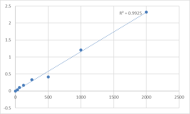
The analytical performance parameters warrant direct statement. The kit achieves a limit of detection of 10 pg/mL and operates across a linear detection range of 31.2–2000 pg/mL. This range captures both the low basal WNT7A concentrations present in healthy tissue and the elevated levels associated with pathological conditions, without requiring separate low-sensitivity and high-sensitivity kit variants for different sample types. Generic WNT7A ELISA kits that bottom out at 50 pg/mL miss the subtle expression shifts that characterize early-stage disease progression or partial pharmacological inhibition—precisely the data range that separates a statistically significant treatment effect from an ambiguous trend line.
Sample compatibility is specified as cell culture supernatants, serum, plasma, and other biological fluids. This breadth matters because WNT7A is a secreted protein that functions in extracellular signaling; its biologically relevant pool is the one present in the conditioned medium, the interstitial fluid, or the circulation, not the intracellular pool detectable by western blot in whole-cell lysate. A kit that quantifies only cell lysates is measuring WNT7A that has not yet been secreted and is therefore not yet biologically active. KTE60019 measures the fraction that actually reaches target cells.
The assay workflow follows the standard sandwich ELISA format with a working time of 3–5 hours, depending on operator experience. The kit ships on gel packs with blue ice and stores at 2–8°C. Components include the pre-coated microplate, WNT7A standard, detection antibody, streptavidin-HRP, standard diluent, assay buffer, HRP substrate, stop solution, wash buffer, and plate covers. The protocol emphasizes standard ELISA disciplines: allow all reagents to warm to room temperature for at least 30 minutes before opening, use fresh pipette tips for each sample and standard to avoid cross-contamination, run samples and standards in duplicate or triplicate, and keep unused wells desiccated at 4°C in the sealed bag provided. These are not exotic precautions. They are the ordinary courtesies that any quantitative immunoassay demands, stated transparently.
The product page indicates zero publications citing KTE60019 at present. This is not a liability for a recently launched kit whose design specifications align with the analytical demands of Wnt signaling research. Every widely adopted ELISA kit began with zero citations. What drives adoption is a combination of antibody specificity, detection sensitivity, sample type flexibility, and protocol reproducibility—all of which are documented in KTE60019's technical specifications. The kit enters a research landscape where Wnt signaling is among the most intensively studied pathways in developmental biology, oncology, and regenerative medicine, and where reliable quantification of individual Wnt ligands has lagged significantly behind quantification of downstream pathway activation markers such as β-catenin nuclear translocation or TCF/LEF reporter activity.
The broader biological context makes reliable WNT7A quantification increasingly difficult to ignore. In oncology, WNT7A overexpression drives breast cancer metastasis and colorectal cancer progression, while loss of Wnt7a in mouse models increases lung tumorigenesis through reduced cellular senescence. A 2024 study identified Wnt7a as a novel upstream inducer of β-catenin-independent tumor-suppressive cellular senescence in lung cancer, demonstrating that Wnt7a-null mice exhibit increased tumor burden not because of altered apoptosis or autophagy, but because senescent cell surveillance is impaired. In head and neck squamous cell carcinoma, WNT7A's role and molecular mechanisms remain incompletely characterized, creating an active research frontier that demands quantitative protein measurement. In colorectal cancer, WNT7A expression in tumor tissues and cell lines correlates with proliferation and disease progression. In reproductive biology, WNT7A is essential for uterine smooth muscle patterning and the maintenance of adult uterine function, and its dysregulation is implicated in infertility and reproductive tract abnormalities.
For the cancer biologist quantifying WNT7A in patient-derived xenograft models, the developmental biologist measuring morphogen gradients in organoid cultures, or the immunologist investigating Wnt-mediated regulation of tissue-resident stem cell populations, the ability to measure WNT7A protein concentration directly and specifically is not an optional upgrade. It is the measurement that converts a gene expression correlation into a protein-level mechanism. KTE60019 provides that measurement in a sandwich ELISA format that distinguishes WNT7A from its closest homologs, detects it at single-digit picogram-per-milliliter concentrations, and accepts the sample types that actually contain the biologically active secreted protein fraction.
Explore specifications, download the protocol, and place your order here: https://www.abbkine.com/product/human-protein-wnt-7a-wnt7a-elisa-kit-kte60019/





