




The Iron Secret Hiding in Your Autophagy Assay: Why Quantifying NCOA4 Changes How You Read Ferroptosis, Iron Toxicity, and Cancer Growth — And How KTE61342 Finally Puts a Number on It
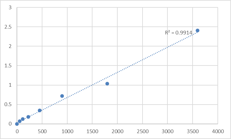
If you've been publishing ferroptosis or iron-metabolism papers recently, you've definitely written the sentence "NCOA4 mediates ferritinophagy" — but chances are you've been measuring it the old-school way: a 64 kDa band on a 10% gel, normalized to actin, with the implicit assumption that "it's there" means the pathway is on. That assumption is exactly where the reproducibility cracks start. NCOA4 (Nuclear Receptor Coactivator 4, alias ARA70/EBP70, UniProt: Q13770, Gene ID: 8021) is a ~624-aa, ~64–70 kDa multi-domain scaffolding protein that wears two identities: it was born as an androgen receptor coactivator (hence the name), but it has been reborn in the last decade as the canonical cargo receptor for selective autophagy of ferritin — aka ferritinophagy — the process that decides whether intracellular iron stays safely locked in its storage vault or gets released as labile Fe²⁺ that can fuel Fenton chemistry and ferroptotic cell death. The Human Nuclear Receptor Coactivator 4 (NCOA4) ELISA Kit (KTE61342) from Abbkine is the reagent that turns this pivotal scaffold into a calibrated, plate-readable ng/mL (or pg/mL) readout, so your iron-metabolism, ferroptosis-sensitization, and muscle-atrophy stories rest on interpolation from a standard curve — not a densitometry guess at 64 kDa.
NCOA4 in One Paragraph: From AR Coactivator to Ferritin's Executioner
NCOA4 was cloned in the late '90s as a 70 kDa protein that binds the androgen receptor (AR) N-terminal AF-1 domain and enhances AR-dependent transcription in a ligand-dependent fashion — hence ARA70. It carries a coiled-coil U-box–like scaffold that mediates both protein–protein interactions and dimerization/tetramerization, and it appeared in early ChIP/pull-down screens as a transcriptional co-regulator that could be broadly nuclear. But the field's center of gravity shifted when Mancias, Kimmelman, and colleagues demonstrated that NCOA4 is the cargo receptor that selectively recognizes ferritin (specifically the Ferritin Heavy Chain, FTH1) and delivers it to the autophagosome via its LIR (LC3-interacting region) motif.
The chain of logic is now canonical:
Condition What NCOA4 Does
Iron deficiency / high demand (hepcidin low) NCOA4 ↑, binds FTH1 → shuttles ferritin to autophagosomes → ferritinophagy → labile iron released → fuels heme/Fe/S cluster biosynthesis
High iron / hepcidin high NCOA4 suppressed (indirectly via mTORC1/4EBP1/iron-sensing axes) → ferritin preserved → iron stays vaulted
Ferroptosis sensitization Excess ferritinophagy → labile iron spike → •OH via Fenton → GPX4-overwhelmed → ferroptotic death
Cancer — paradox NCOA4 can either (a) supply iron for proliferative metabolism, or (b) trigger ferroptosis if tumor's GPX4/FSP1 system is weak; context is everything
NCOA4 also moonlights in NRF2 coactivation and circadian CRY2 signaling, and loss-of-function work implicates it in muscle atrophy, renal proximal tubule iron handling, and Friedreich's ataxia–adjacent iron neurotoxicity, making it one of the most cross-disciplinary "specialized scavenger" proteins in the modern metabolome.
Why a Sandwich ELISA for NCOA4 — And Why Gel-Only Fails You on the Ferritinophagy Claim
NCOA4 is largely cytosolic/soluble, runs ~64–70 kDa (sometimes a 55 kDa shorter isoform), and shifts its mobility depending on post-translational modifications (phosphorylation, ubiquitination) and association with FTH1 complexes. That creates three practical problems:
- The "64 kDa zone" is crowded — particularly in muscle, kidney, and liver lysates where actin-binding and chaperone proteins proliferate. A single antibody can catch a cross-reactive band that looks like NCOA4 but isn't.
- Ferritinophagy changes NCOA4's solubility/turnover: when autophagy is induced, NCOA4–FTH1 complexes can traffic or get degraded along with the cargo — so your "band intensity" confounds amount present with fraction degraded during the assay window.
- Replicates & time courses (Erastin/RSL3 time courses, hepcidin time courses, iron-chelator panels) demand a plate-based number with CVs you can defend.
The KTE61342 kit uses the field-standard two-site sandwich ELISA:
- Microplate pre-coated with a capture antibody specific for human NCOA4.
- Standards (recombinant human NCOA4) + samples — tissue homogenates, cell lysates, cell culture supernatants/lysates, other biological fluids — added → NCOA4 binds.
- Wash → biotinylated anti-NCOA4 detection antibody (different epitope) → Streptavidin–HRP → TMB → color ∝ bound NCOA4.
- Stop → read 450 nm → interpolate NCOA4 concentration from the standard curve.
Representative performance envelope for this kit family:
Parameter Typical KTE61342-class spec
Target Human NCOA4 / ARA70 (UniProt Q13770, Gene ID 8021)
Format 96-well sandwich ELISA, pre-coated capture
Detection Biotin-Ab → SA-HRP → TMB, 450 nm
Dynamic Range 0.156 – 10 ng/mL (7-point standard)
Sensitivity / LOD ~0.05–0.10 ng/mL
Intra-Assay CV < 7–8%
Inter-Assay CV < 10–12%
Samples Tissue homogenates, cell lysates, culture supernatants, serum/plasma (exploratory)
Assay time ~3–5 hours
(As always, anchor your exact dilution scheme and lot-specific recovery to the shipped Abbkine datasheet/CoA.)
Where Quantifying NCOA4 Protein Actually Moves the Story
- Ferroptosis / Lipid Peroxidation Screens
The canonical ferroptosis inducers — Erastin (system Xc⁻ inhibitor), RSL3 (GPX4 covalent inhibitor), FIN56, imidazole ketones — work through or parallel to the labile-iron pool. NCOA4 is the genetic rheostat of that pool:
• NCOA4 KO / siRNA → ferritin not degraded → labile iron ↓ → RESISTANCE to RSL3/Erastin
• NCOA4 OE / hepcidin-low conditions → ferritinophagy ↑ → labile iron ↑ → SENSITIZATION
Measuring NCOA4 (ng/mg total protein, BCA) alongside FTH1, GPX4, ACSL4, and lipid-ROS (C11-BODIPY/ Liperfluo) closes the mechanistic chain: you're not just showing "cells died," you're showing the iron-delivery conduit scaled with the death.
- Iron-Overload Pathology & Hereditary Hemochromatosis Modifiers
Why do some HFE-mutation carriers load iron aggressively while others don't? One layer is how efficiently ferritinophagy clears excess stored iron when demand spikes — and NCOA4 protein levels (in liver biopsy lysates or, exploratorily, in peripheral markers) become part of the penetrance story.
- Kidney: Renal Proximal Tubule Injury & Nephrotoxins
The proximal tubule reabsorbs and processes ~95% of filtered protein; it's also exquisitely sensitive to hemoglobin/myoglobin-derived iron, cisplatin (which induces ferroptosis), and heme-iron cycling. NCOA4 induction here is protective in some windows (releasing iron for repair) and catastrophic in others (fueling tubular necrosis). Quantifying it in brush-border / cortical lysates gives you the ferritinophagy axis number.
- Muscle Atrophy & Duchenne-Related Cachexia
Chronic illness, disuse, and dystrophin-loss models show iron dysregulation in atrophying muscle. NCOA4-mediated ferritinophagy can release labile iron that drives proteolysis and exacerbates wasting; measuring NCOA4 in gastrocnemius/quadriceps lysates (normalized to mg protein) alongside FTH1, 4HNE, DCF, and MuRF1/atrogin-1 builds a quantitative iron-toxicity limb.
- Neurodegeneration (Friedreich's Ataxia, Parkinsonian Iron Accumulation)
Frataxin deficiency → mitochondrial Fe–S deficit → cytosolic labile iron ↑ → NCOA4 may attempt compensatory ferritinophagy → if GPX4 system is stressed, neurons tip into ferroptosis-like damage. iPSC-derived dopaminergic or cerebellar neurons benefit from NCOA4 quantification as the "iron-release valve" readout.
- CRISPR/AAV Validation
Editing NCOA4? Report % NCOA4 protein remaining ± SEM from a calibrated curve, normalized to mg total protein (BCA) — and co-show FTH1 levels (often reciprocally regulated by degradation), labile iron (Calcein-AM quenching), and lipid-ROS. Reviewers consistently prefer this triad over a lone "band fainter."
A Minimal Prep Blueprint (NCOA4 Is Cytosolic — Treat It Like a Soluble Node)
• For tissues (liver, kidney, muscle, brain): homogenize cold in 50–100 mM Tris, pH 7.4, 150 mM NaCl, 0.5–1% Triton X-100/NP-40 + protease inhibitors + 5–10 mM deferoxamine (optional, to stabilize iron-sensitive complexes during prep), clarify 12,000–16,000 ×g, 15 min, 4°C → supernatant is your NCOA4 pool.
• For whole-cell lysates: same detergent buffer works; spin, keep cold, BCA.
• Express as ng NCOA4 / mg total protein.
• Warm kit reagents ≥ 30 min RT before opening; protect TMB from light; stop uniformly; read 450 nm promptly; run the full standard curve on every plate — iron-protein recoveries can vary with hemolysis/metal adjuvants, and the curve is your insurance.
The Bottom Line
NCOA4 is the 64 kDa scaffold that sits at the hinge between androgen signaling, nuclear coactivation, and — most urgently — the autophagic release of vaulted iron via ferritinophagy. If your experiment touches ferroptosis, iron-chelation, hepcidin biology, muscle wasting, or renal tubular iron injury, NCOA4 isn't optional background — it's the conduit that decides whether iron stays safe or becomes a Fenton catalyst. The Human Nuclear Receptor Coactivator 4 (NCOA4) ELISA Kit — KTE61342 from Abbkine gives you the right architecture to measure it: pre-coated capture → biotin detection → HRP–TMB → 450 nm → ng/mL, over a 0.156–10 ng/mL working range, in a ~3–5 hour workflow that scales across genotypes, drug panels, and tissue cohorts without chaining you to a gel rig.
Product Reference: KTE61342 – Human Nuclear Receptor Coactivator 4 (NCOA4) ELISA Kit
Learn more and order: https://www.abbkine.com/product/human-nuclear-receptor-coactivator-4-ncoa4-elisa-kit-kte61342/
(For Research Use Only; not for diagnostic procedures in humans.)





