


The Executioner You Can't Afford to Misread: Why Your Bax Antibody Choice Dictates Every Apoptosis Figure's Fate — And How Abbkine's ABP55948 Delivers
BAX (Bcl-2-associated X protein) is the one pro-apoptotic switch the entire intrinsic pathway converges on — and the protein that most "apoptosis Westerns" take for granted until the reviewer asks the uncomfortable question: how do you know that 21 kDa band is actually Bax, and not a Bcl-2 cross-reactive shadow? Every first-year cell biology student memorizes the dogma: Bax + Bak = mitochondrial outer membrane permeabilization (MOMP) → cytochrome c release → caspase-3/-7 activation → controlled demolition of the cell. But the gap between "the pathway diagram" and a publishable, reviewer-proof figure is where your antibody does the heavy lifting — or quietly sabotages you. The Bax Polyclonal Antibody (ABP55948) from Abbkine is built for labs that need more than a band — it's an affinity-purified, rabbit-derived IgG validated across Western blot, IHC on paraffin (IHC-P), immunofluorescence (IF/ICC), and ELISA, so your Bax signal survives both the confocal microscope and the peer review.
BAX in One Clean Paragraph: Cytosol → Mitochondria → The Point of No Return
BAX (UniProt: Q07812, Gene ID: 581) is a 21–23 kDa (~21 kDa for the dominant Bax-α isoform), mostly α-helical protein with three Bcl-2 homology domains (BH1–BH3) and a C-terminal transmembrane anchor that keeps it engaged with — or ready to insert into — the mitochondrial outer membrane (MOM). In a healthy, unstressed cell, the vast majority of Bax is cytosolic, held in soluble monomers/touching the outer membrane lightly via interactions with 14-3-3 (bound/phospho-regulated) and other chaperones. The moment intrinsic stress hits — DNA damage (p53↑), ER stress, growth factor withdrawal, ROS overload, or direct BH3-only activator (tBid, BIM, PUMA) — Bax undergoes a conformational flip: the BH3-exposed, membrane-insertion-ready form emerges, Bax translocates, oligomerizes into lipid-embedded pores, and the MOM springs a leak. Cytochrome c, Smac/DIABLO, Omi/HtrA2 spill into the cytosol. Apoptosome assembles. Caspases fire. Game over.
The biological decision-point every cancer therapist exploits is brutally simple:
Bax (pro-death) : Bcl-2/Bcl-XL (anti-death) ≈ the rheostat that decides whether the cell lives or dies.
Push Bax up → death. Push Bcl-2 up → survival. The Bax/Bcl-2 ratio is so reproducible it has been used as a prognostic index in chemoresistance, lymphoma grading, and treatment-response stratification.
Why ABP55948: The Polyclonal Advantage — Done Right
Monoclonals are elegant, but a well-made rabbit polyclonal against Bax earns its place in three very specific situations:
Challenge Why ABP55948's Approach Wins
Conformational complexity (soluble monomer vs. membrane-inserted oligomer vs. cleaved/modified forms) Multi-epitope recognition across the internal/spanning regions of Bax improves the odds of catching Bax regardless of subtle conformational masking — especially in IHC-P where fixation/embedding can freeze the protein in a less accessible state
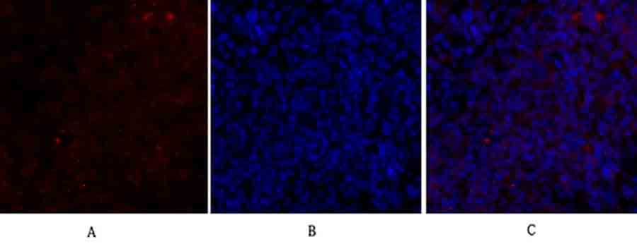
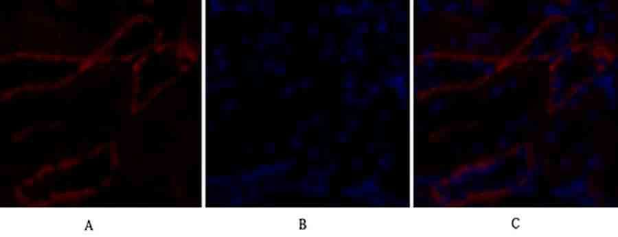
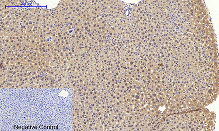
Detection flexibility One affinity-purified reagent spans WB (21 kDa sharp band), IHC-P (granular cytoplasmic → mitochondrial pattern, intensifying at apoptotic foci), IF/ICC (translocation tracking), and ELISA capture
Signal robustness for low-Bax basal conditions Many unstressed cells express Bax but keep it at "quiet" levels — a polyclonal's aggregate affinity can preserve signal where a single-clone's epitope might be borderline
The product specifications you'll cite for ABP55948 cluster around:
• Host / Isotype: Rabbit IgG, polyclonal, peptide-affinity-purified
• Immunogen: Synthesized peptide derived from human Bax (internal/spanning regions — not just the extreme N- or C-tail, which can over-expand phylogenetic cross-reactivity)
• Reactivity: Human, Mouse, Rat (Bio-informatics homology also supports broader vertebrate cross-reactivity in many protocols)
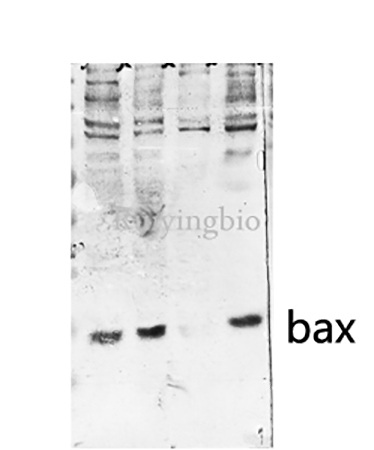
• Observed MW: ~21 kDa (Bax-α), sometimes a fainter ~24 kDa if Bax-β pops
• Applications & typical dilutions:
• WB: 1:500 – 1:2,000
• IHC-P (FFPE): 1:100 – 1:300 (antigen retrieval usually required)
• IF/ICC: 1:100 – 1:1,000
• ELISA: 1:10,000 – 1:20,000 (capture/detection sandwich setups)
• Format: Liquid in PBS, pH 7.4, 50% glycerol, 0.5% BSA, 0.02% sodium azide
• Storage: -20°C, avoid freeze–thaw cycles
The One Thing Everyone Gets Wrong About Bax Westerns (And How to Fix It)
The Mistake: Quantifying Bax Total Protein as "Apoptosis Occurred"
Bax going up is necessary but not sufficient proof of MOMP. Bax can accumulate and still sit cytosolic if the insertion/oligomerization step is blocked (Bcl-2/Bcl-XL sequestration, VDAC2 capping, Pin1/IBRDC2 regulation). The cleanest Bax experiment therefore pairs:
Readout What It Proves
Total Bax (ABP55948 WB) "Bax expression changed (p53-driven? transcriptional?)"
Bax translocation (IF/ICC or subcellular fractionation WB: Bax in 10k-mito pellet vs. cytosol) "Bax actually went to the membrane"
Cytochrome c release / cleaved caspase-3 / Annexin V "MOMP happened and the execution proceeded"
Bcl-2 / Mcl-1 / Bcl-XL The counter-balance that contextualizes why Bax did or didn't fire
The Fix: Your 21 kDa Band Should Look Like This
• Lane: 20–30 µg whole-cell lysate, 12–15% Tris-glycine gel (Bax runs crisp at ~21 kDa, just below the Actin 42 kDa but well above the crowded 14 kDa region if you run it right).
• Positive control: STS (staurosporine 1 µM, 4–6 h) or etoposide (50 µM, 24 h) treated HeLa/HEK293/ Jurkat — this should give you a strong, clean 21 kDa band and nuclear condensation you can brag about in the IF panel.
• Negative control: Untreated cells + secondary-only (confirms no non-specific ~21 kDa ghost).
• Normalize to β-actin, GAPDH, or vinculin — not to Bcl-2 (they reciprocally regulate, so it's circular logic).
Where ABP55948 Earns Its Keep Across Fields
- Cancer chemotherapy & drug-resistance screens
If your compound is supposed to "activate the intrinsic pathway," Bax is your North Star:
• WB: does total Bax rise (p53-on)?
• IF/IHC: did Bax leave the cytosol and pile onto mitochondria?
• ELISA format (sandwich): can you screen 48 conditions in a plate?
BH3 mimetics (venetoclax, ABT-737, S63845, MCL-1 inhibitors) live or die by Bcl-2/Bcl-XL/Mcl-1 neutralization → Bax/Bak liberation → death; your antibody needs to survive the quantification.
- Neurodegeneration & ischemia–reperfusion injury
Hippocampal CA1, substantia nigra, cortical neurons — Bax translocation is the structural commitment to neuronal death after glutamate excitotoxicity, ROS, or cardiac arrest-reperfusion. IHC-P on perfused brain (FFPE) with ABP55948 gives you the granular-to-mitochondrial Bax pattern across the surviving vs. dying zones; IF on primary neurons lets you co-label with TOM20/VDAC to prove co-localization.
- Immune homeostasis & lymphocyte apoptosis
Activation-induced cell death (AICD), negative selection in thymus, Fas/CD95 crosstalk with intrinsic amplification — all route through Bax. Tracking Bax in thymocyte subpopulations or activated T blasts (often low total protein per cell, which is why polyclonal's signal depth helps) is a standard.
- Metabolic stress & β-cell apoptosis
Cytokine (IL-1β + IFNγ) toxicity in human islets, palmitate-induced lipoapoptosis, or hyperglycemia-driven ROS → Bax is the effector that blows the β-cell. Quantifying it in human islet lysates (precious material) benefits from a high-affinity, affinity-purified detection rather than a marginal clone.
- CRISPR/siRNA validation
Knocking BAX out? Report % Bax protein remaining (21 kDa) ± SEM from a calibrated lane, and show the functional payoff: cleaved PARP ↓, cytochrome c retention, Annexin V ↓ — exactly the triplet reviewers want.
A Quick Prep/Controls Checklist
• Lysis: RIPA or Tris-Triton (0.5–1% NP-40) + protease inhibitors, cold, homogenize quickly.
• Fractionation (optional but impressive): 600 ×g (nuclei) → 10,000 ×g 15 min (crude mito-enriched pellet = look for Bax enrichment vs. cytosolic supernatant).
• Gel: 12–15% resolving gel for a tight ~21 kDa band. Transfer: PVDF often improves low-kDa recovery.
• IHC-P retrieval: Heat-mediated (pH 6.0 citrate or pH 9.0 EDTA) — Bax's internal epitopes usually survive formalin well once retrieved.
• Dilution: Start around 1:1,000 for WB (the 1 mg/mL stock goes far), tweak ± to your signal/noise.
• Store at -20°C, avoid repeated freeze–thaw of the stock (aliquot 10–20 µL working volumes if you use it daily).
The Bottom Line
Bax is the pro-apoptotic executioner your cancer drug tries to unleash, your ischemic neuron tries to suppress, and your Western blot pretends is "obvious." But obvious doesn't survive peer review — specificity, subcellular translocation evidence, and a clean 21 kDa band normalized to a non-Bcl-family housekeeper do. The Bax Polyclonal Antibody (ABP55948) from Abbkine gives you that evidence across the platforms that matter: WB (~21 kDa), IHC-P, IF/ICC, and ELISA, in a rabbit IgG, peptide-affinity-purified format you can trust not to confuse Bax with its anti-apoptotic cousins.
Product Reference: ABP55948 – Bax Polyclonal Antibody
Learn more and order: https://www.abbkine.com/product/bax-polyclonal-antibody-abp55948/
(For Research Use Only; not for diagnostic procedures in humans.)


