




The Background You Cannot Afford to Leave Unchecked: How a Single Serum Vial Decides the Fate of Your Most Critical Immunoassays
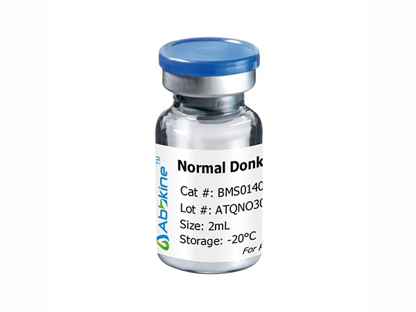
Every immunoassay protocol ever written contains a step so routine, so seemingly pedestrian, that it is performed almost without thought. You block your membrane, your tissue section, your ELISA plate with BSA or non-fat dry milk, incubate, wash, and trust that the background will remain low enough to distinguish signal from noise. But there is a quiet, persistent limitation in this approach that only reveals itself when the experiment fails. The hydrophobic patches on your Fc receptors are not uniformly occupied by albumin molecules. Endogenous immunoglobulins in your tissue are not inert. Non-specific binding sites on your extracellular matrix are not fully saturated. And the result, when you finally sit down at the microscope or the imager, is mottled background, cytoplasmic haze, and a negative control that looks distressingly positive. The block was never a formality; it was the foundation upon which your entire detection system rested. Using a non-immune serum from the same host species as your secondary antibody is the most biochemically appropriate way to build that foundation correctly. Abbkine's Normal Mouse Serum (BMS0070) is engineered precisely for this purpose, refined from collection to formulation to eliminate the common failure points that degrade immunoassay reproducibility. And with six peer-reviewed publications already citing it—including a study in Colloids and Surfaces A (IF 5) that deployed the product during the investigation of peptide-mediated targeting of hepatocellular carcinomas—the research community has begun to take notice.
What sets Abbkine's Normal Mouse Serum apart, before any discussion of dilution factors or blocking times, is its provenance—a detail that most serum datasheets obscure under the vague term "normal." and that, misinterpreted, introduces confounding immunological variables that no amount of optimized protocol can later correct. The serum is processed from blood collected exclusively from non-immunized normal adult mice, a specification that carries real immunological weight. Serum from immunized or even environmentally challenged mouse colonies can carry elevated titers of polyclonal antibodies against common laboratory antigens—bovine serum albumin, casein, plant lectins from standard chow—that cross-react with blocking reagents, detection antibodies, and target proteins in unpredictable ways. Younger mice possess an immature complement system and a distinct serum protein profile, with immunoglobulin levels that differ substantially from the adult. Serum from non-immunized adult mice contains the full, balanced complement of serum proteins—albumin, globulins, complement factors, protease inhibitors, and the normal polyclonal IgG repertoire—that collectively saturate the diverse non-specific binding sites present in fixed tissue and on assay substrates. Crucially, the endogenous mouse IgG in the serum occupies Fc receptors that would otherwise capture your goat anti-mouse or donkey anti-mouse secondary antibody, generating the characteristic "membrane-edge background" or "fibroblast-shaped artifacts" that IHC laboratories have learned to recognize and dread.
The preparation workflow through which BMS0070 passes after collection reveals specifications that are not cosmetic but functionally consequential. First, hemolysis is minimized by employing proprietary techniques during the blood collection and serum extraction processes. Hemolyzed serum is not a cosmetic problem; it is an analytical liability. Free hemoglobin released from lysed red blood cells possesses potent pseudoperoxidase activity that catalyzes the conversion of DAB substrate in HRP-based detection systems, producing insoluble brown precipitate at sites indistinguishable from genuine antigen localization—a phenomenon that has led more than one graduate student to excitedly report "nuclear staining" that was, in reality, hemoglobin-catalyzed artifact nucleation. By controlling hemolysis at the collection stage, BMS0070 removes a background variable that most serum manufacturers fail even to acknowledge.
Second, the serum undergoes lipid extraction to improve clarity. This optical specification translates directly into operational performance in fluorescence-based detection modalities—immunofluorescence, confocal microscopy, super-resolution imaging—where residual lipid micelles scatter excitation light and produce the punctate autofluorescence artifacts that survive all subsequent washes and photobleaching corrections. A blocking reagent that introduces fluorescent noise is not a blocker; it is a confound. Third, the extracted serum is dialyzed against 10 mM sodium phosphate, 0.15 M sodium chloride, pH 7.2, a buffer composition that precisely matches phosphate-buffered saline and eliminates the osmotic shock that can occur when a high-protein serum solution contacts delicate tissue architecture. And fourth, the product is supplied as a ready-to-use liquid solution rather than as a lyophilized powder that requires reconstitution, centrifugation, sterile filtration, and concentration verification before use. The liquid presentation eliminates the rehydration step that lyophilized sera demand—a step that, when performed incompletely or with non-sterile water, introduces particulate background, microbial contamination, and protein concentration variability across aliquots that no amount of subsequent dilution can correct.
The recommended working dilutions for BMS0070 reflect the protein concentration of whole serum rather than exaggerated marketing claims. The product documentation specifies suggested starting dilutions of 1:10–1:20 (equivalent to 5–10% v/v) for immunohistochemistry on paraffin-embedded tissue and immunofluorescence or immunocytochemistry, and 1:100 for ELISA. These are realistic numbers. Normal serum diluted to 5%–20% (v/v) in PBS is the empirically validated range that the immunochemistry literature has converged upon over decades—concentrations below this range risk incomplete blocking of abundant non-specific binding sites, while concentrations above this range increase solution viscosity, complicate washing, and can elevate background through excess immunoglobulin deposition on the tissue section. The 1:100 ELISA recommendation reflects the lower non-specific binding burden of polystyrene plate surfaces compared to the complexity of fixed tissue, and the fact that many ELISA protocols incorporate detergent-based washing steps that serum blocking alone cannot replace. Abbkine specifies that optimal working dilutions should be determined experimentally by the investigator, a statement of biochemical honesty rather than protocol evasiveness. Tissue type, fixation method, antibody affinity, and detection system sensitivity all influence the optimal blocking concentration, and no manufacturer can predict these variables for every experiment every user will perform.
The dual functionality of Normal Mouse Serum—as both a blocking reagent and a negative control—deserves explicit emphasis because it distinguishes serum from every chemically defined blocking alternative on the market. As a blocking reagent, BMS0070 is most effective when the serum matches the host species of the labeled secondary or tertiary antibody. When your detection system employs goat anti-mouse IgG, the goat IgG present in normal goat serum saturates the Fc receptors and conserved-sequence binding sites that would otherwise capture your secondary antibody; when your detection system employs goat anti-rabbit IgG and your tissue is mouse, normal mouse serum blocks the endogenous mouse immunoglobulins that would cross-react. The blocking specificity is not species-agnostic; it follows the secondary antibody. As a negative control, normal serum from the same host species as the primary antibody—diluted to the same total protein concentration as the primary antibody working solution—provides the most biochemically accurate assessment of non-specific primary antibody binding. Unlike buffer-only controls that reveal only the background from secondary antibody binding to tissue, a normal serum control at matched immunoglobulin concentration reveals the background from non-specific primary antibody Fc region binding, hydrophobic interaction, and charge-based adsorption, which is the background that actually competes with specific antigen-antibody signal. For laboratories using mouse monoclonal primary antibodies—and mouse remains the dominant host species for monoclonal antibody production—the availability of a validated normal mouse serum for both blocking and negative control experiments eliminates the need to source, validate, and optimize separate reagents for these two analytically distinct functions.
The published literature provides the independent validation that no manufacturer's in-house performance characterization can substitute. Six publications currently cite BMS0070, spanning journals and application types that test different dimensions of the serum's performance. One of those studies examined how peptide-mediated targeting of human hepatocellular carcinomas could be enhanced through composition-engineered protein corona engineering on gold nanoparticles, an application that required blocking conditions producing near-zero non-specific binding in nano-bio interface assays where even picomolar non-specific protein adsorption confounds quantitative interpretation. Another publication, appearing in Molecular Medicine Reports (IF 2), evaluated formaldehyde-induced apoptosis of BALB/c mouse bone marrow cells through the PTEN/PI3K/Akt signal transduction pathway, relying on BMS0070 for immunohistochemical blocking in a context where formalin-fixed paraffin-embedded mouse tissue sections present the endogenous mouse IgG that generates the most notorious source of background in mouse-on-mouse IHC staining. A 2026 Abbkine technical blog noted that this serum has accumulated over 4,000 product page views, indicating sustained interest from the research community. Every published figure that lists BMS0070 in its methods section represents an independent laboratory that processed its own tissues, applied its own primary antibodies, detected with its own secondary reagents, and submitted its own data for peer review with this specific serum mentioned as the blocking and control reagent. The aggregate validation from six independent laboratories, operating in cancer biology, toxicology, and nanomaterials research contexts that demand precisely the blocking specificity that Normal Mouse Serum provides, is more informative about real-world performance than any manufacturer's certificate of analysis.
The storage and stability specifications reward careful reading because they determine whether the reagent in the freezer today performs identically to the same reagent used three months from now. BMS0070 stores at 2–8°C for short-term use, or can be aliquoted and stored at -20°C or below for long-term storage, with the explicit instruction to avoid repeated freezing and thawing. This is not a generic disclaimer. Freeze-thaw cycling of whole serum promotes the formation of cryoprecipitate—aggregated immunoglobulins and lipoprotein complexes that precipitate upon thawing and, once formed, do not fully resolubilize upon vortexing or warming. These microscopic protein aggregates deposit on tissue sections during the blocking incubation, producing the particulate background artifacts that confound high-magnification imaging and persist through mounting medium application. Aliquoting the serum into single-use volumes upon the first thaw eliminates this degradation pathway entirely, preserving the homogeneous protein composition that the manufacturer validated. The formulation includes 0.05% sodium azide as a preservative, a concentration sufficient to inhibit microbial growth during long-term storage without reaching the levels (≥0.1%) that inhibit horseradish peroxidase enzymatic activity when residual azide carries over from the blocking step into the HRP-conjugated secondary antibody incubation. The product ships on gel packs with blue ice, maintaining the cold chain from the manufacturer's warehouse to the laboratory receiving dock. Available package sizes include 2 mL and 10 mL vials, with bulk sizes available upon request.
The broader immunological context makes the case for proper blocking reagent selection increasingly difficult to dismiss as multiplexed, high-sensitivity, and super-resolution imaging technologies proliferate. Tyramide signal amplification (TSA)-based multiplexed immunohistochemistry deposits covalent fluorophore-tyramide conjugates at the precise site of HRP activity, permanently labeling the tissue section with fluorophores that cannot be chemically removed without compromising epitope integrity. Any non-specific HRP deposition during the amplification step—including HRP bound to endogenous mouse immunoglobulins that were not adequately blocked, or HRP captured by pseudoperoxidase-active hemoglobin from hemolyzed blocking serum—produces a permanent signal that accumulates error across sequential staining, stripping, and reprobing cycles. In cyclic immunofluorescence methods such as CyCIF, CODEX, and 4i, where the same tissue section undergoes 10–40 sequential rounds of primary antibody incubation, secondary antibody detection, imaging, and antibody stripping, the cumulative probability of blocking failure across cycles approaches certainty unless the blocking reagent performs consistently across every round. In super-resolution microscopy, where single-molecule localization precision approaches 10–20 nm, background fluorescence from inadequately blocked non-specific binding sites directly degrades the localization accuracy that defines the technique's value. In every one of these experimental contexts, proper blocking reagent selection—using normal serum from the species matching the detection antibody, at the empirically determined optimal dilution, with full awareness of the hemolysis, lipid content, and protein composition of the serum—is the single most controllable variable separating publication-quality images from troubleshooting sessions that consume afternoons and erode research budgets.
For the graduate student whose IHC images show brown precipitate everywhere, the core facility technologist who processes hundreds of tissue sections per month and cannot afford a single failed blocking step, the immunology laboratory whose multiplexed tissue imaging experiment depends on the cumulative integrity of 40 sequential blocking and staining rounds, the toxicology researcher investigating apoptosis pathways in formalin-fixed mouse bone marrow sections, the nanomaterials scientist whose nanoparticle cellular uptake assays demand near-zero non-specific protein background, and the investigator who simply wants the negative control slide to look like a negative control slide—meaning no brown staining, no fluorescent haze, no particulate debris, no binding artifacts that survive peer review—BMS0070 provides a blocking and control reagent whose source animal health status, hemolysis-minimized collection procedure, lipid-extracted clarity, whole-serum protein composition representing the complete mouse immunoglobulin repertoire, PBS-matched dialysis buffer, and six peer-reviewed publications collectively answer the question that the blocking step poses to every immunoassay: will the signal be signal, and will the background be background?
Don't let your next groundbreaking result be lost to non-specific noise. Explore specifications, access the detailed protocol FAQ, and place your order here: https://www.abbkine.com/product/normal-mouse-serum-bms0070/





