




That "Clean 21 kDa Band" in Your G1-Arrest Western Might Be a Ghost—Here's Why a T145-Centered p21/CDKN1A Polyclonal (ABP0108) Is the Only Way To Shut Down the "Is It Really p21?" Reviewer Debate
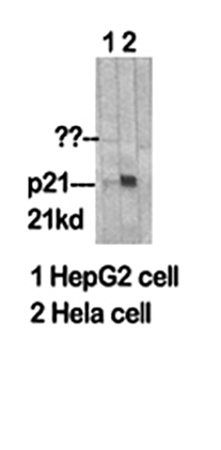
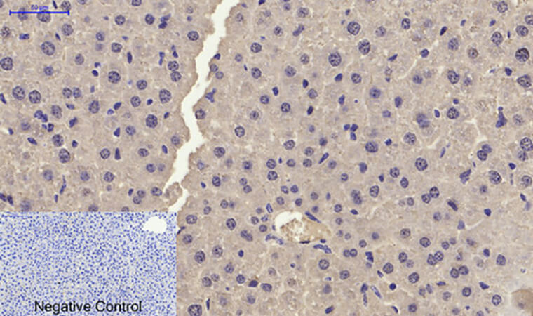
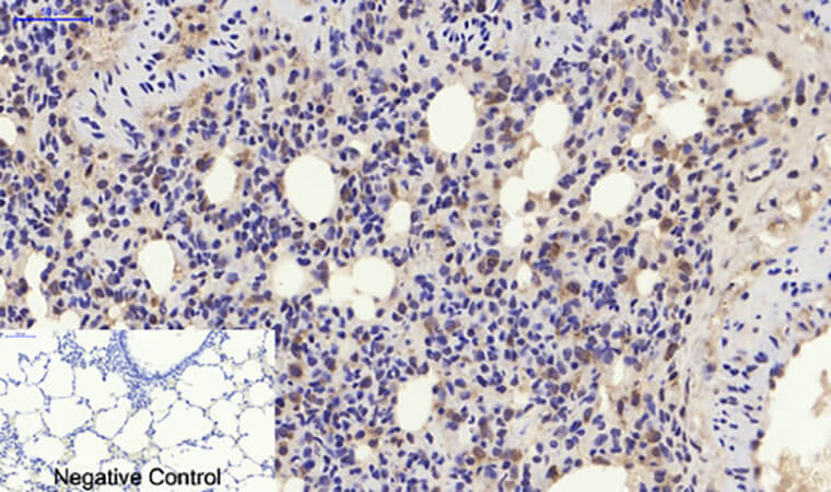
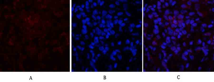
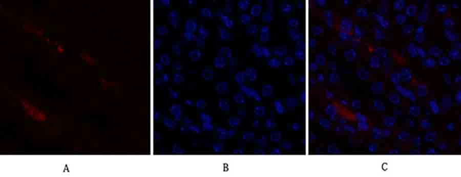
There's a very specific kind of frustration that visits every lab running cell cycle, senescence, or DNA-damage response screens: you've irradiated your cells (or hit them with etoposide/doxorubicin), your p53 Western looks great, your cyclin D1/CDK4 bars are dropping on schedule, and there—at ~21 kDa—is the band you think is p21/CDKN1A (WAF1/CIP1) doing its G1-braking job. But then the revision letter lands with the line that makes you groan: "The authors should provide additional validation (e.g., with a second p21 antibody raised against a distinct epitope, or p21-knockdown control) to confirm the identity of the ~21 kDa signal." And suddenly you realize your "p21" came from a generic cyclin-dependent kinase inhibitor panel antibody that was raised against a GST-fusion covering the PCNA-binding region—which also happens to cross-react with a few PCNA-associated or caspase-cleaved fragments that run distressingly close to 18–21 kDa.
p21/CDKN1A Is NOT a "Bonus Marker"—It's the p53-Dependent Gatekeeper That Decides Whether Your Cells Arrest or Die
CDKN1A (p21, WAF1, CIP1 — UniProt P38936, Gene ID 1026) is the canonical p53 transcriptional target that binds to and inhibits cyclin E/A–CDK2 and cyclin D–CDK4/6 complexes, enforcing the G1-phase checkpoint in response to DNA damage, oncogenic stress, and a growing list of senescence-triggering cues (telomere erosion, persistent DDR foci, SASP cross-talk). What makes p21 uniquely complicated—and why antibody choice actually matters—is its functional yin-yang duality:
Context What p21 Does Why Your Antibody Choice Matters
Nucleus (canonical) Binds CDK2/4 → G1 arrest, buys time for DNA repair You need an epitope away from the CDK-binding pocket so the band doesn't vanish just because p21 is in a complex
Cytoplasm Binds/modulates caspase-3 and can have anti-apoptotic effects under specific stress Cytoplasmic "sticky" backgrounds can masquerade as p21 if your antibody isn't epitope-purified
Caspase-cleavage zone (D147↓) Cleaved p21 loses CDK-inhibition, can promote CDK2 reactivation A good antibody should still recognize the intact T145 epitope region (pre-cleavage) while not hallucinating random 14 kDa fragments
The bottom line: where your immunogen sits on the p21 sequence determines whether your ~21 kDa band is p21 or a persistence-prone artefact.
Why ABP0108 Is Built Different: The Immunogen Is Centered on T145, Not a Random GST-Fusion
Here's the strategic detail that separates Abbkine's ABP0108 — p21 Polyclonal Antibody from the "just-anti-p21" crowd:
Parameter ABP0108 Specification
Target p21 / CDKN1A / WAF1 / CIP1 (UniProt P38936, Gene ID 1026)
Aliases CDKN1A; CAP20; CDKN1; CIP1; MDA6; PIC1; SDI1; WAF1; melanoma diff.-associated protein 6
Host / Isotype Rabbit · IgG · Polyclonal
Immunogen Synthesized peptide derived from human p21, centered on the non-phosphorylation site T145 (AA range ~111–160 context)
Reactivity Human, Mouse, Rat (H/M/R cross-validated)
Observed MW ~21 kDa (calculated ~18 kDa; runs ~21 kDa on standard SDS-PAGE)
Cellular Localization Nucleus (primary), also reported cytoplasm / mitochondria-associated depending on stress & isoform context
Validated Apps & Starts WB: 1:500–2,000 · IF: 1:50–200 · IHC-P: 1:50–300 · ELISA: ~1:20,000
Format / Conc. Liquid, 1 mg/mL in PBS pH 7.4 + 0.5% BSA + 0.02% NaN₃ + 50% glycerol
Purification Epitope-specific affinity purification from rabbit antiserum → higher specificity than crude IgG fractions
Storage / Ship -20°C, 1-year stable from shipment; centrifuge vial after thawing/before opening; aliquot to avoid repeated freeze–thaw; ships blue-ice gel pack
Status For research use only; not for human/clinical diagnostic use
The differentiator is right there in plain sight: the immunogen is a synthetic peptide centered on the T145 region, not a whole-protein GST-slurry covering the PCNA-binding domain where every Tom, Dick, and Harry kinase/adaptor also hangs out. Epitope-affinity purification further strips away the "background-loving" IgG fraction you get from a raw serum prep. The result is a ~21 kDa nuclear band that actually belongs to p21/CDKN1A.
What Actually Changes in Your Paper When p21 Behaves Instead of Needing Three Paragraphs of Apology
① Your DNA-damage / G1-arrest story becomes causally tight.
Run ABP0108 at 1:1,000–2,000 on an etoposide / IR / hydroxyurea time course with a positive control (p53-wild-type cells like MCF-7, HeLa, U2OS, or HUVEC) and a negative anchor (p53-null cells like H1299, SaOS-2, or p21-siRNA knockdown). When your ~21 kDa band rises on cue and collapses in the knockdown, the reviewer sees evidence—not a hoped-for interpretation.
② Your senescence & SASP panels stop relying on SA-β-Gal alone.
p21 is the effector that holds the senescent growth arrest (while p16/INK4a often joins later). Having a clean IHC-P signal at 1:100–200 on FFPE sections (with EDTA pH 8.0 or citrate pH 6.0 retrieval) lets you map where in the tissue architecture the arrest-program is engaged—not just "β-Gal stained blue somewhere."
③ One vial, three platforms, zero reinvention.
Because it covers WB + IHC-P + IF (+ ELISA-range utility), your lysate screen → xenograft validation → FFPE TMA scoring all ride the same epitope, keeping your Methods compact and your batch-consistency sane.
The Bench Rules That Keep Your ~21 kDa Band Sharp (and Your Friday Night Intact)
p21 is a nuclear, relatively low-abundance CDKI (~18–21 kDa) that spikes post-damage but can easily be obscured by:
• Overloaded total protein (makes every band look "smear-y")
• Insufficient blocking (BSA vs. non-fat milk debate—p21 Westerns often prefer 5% BSA to avoid cross-reacting milk proteins near 20–25 kDa)
• Residual azide inhibiting HRP if you don't wash after the primary
The habit-list that saves you:
• Centrifuge before opening — 1 mg/mL in 50% glycerol means stratification; a 10-sec spin recovers the full concentration from the cap.
• Aliquot on Day 1 (5–10 µL tubes). The -20°C/glycerol format is stable, but repeat freeze–thaw is the #1 cause of IgG aggregation and high nuclear background.
• Run a positive AND a p21-siRNA/p53-null lane once — nothing shuts down the "identity of the ~21 kDa signal" critique faster than a side-by-side where it vanishes.
• WB dilution scouting: start 1:1,000 on a damage-induced lysate (50–80 µg total protein, 10–12% Tris-Glycine gel works nicely for ~21 kDa separation from actin at ~42). If your signal is strong, push to 1:2,000 for a gorgeously clean lane.
• For IHC-P on FFPE: standard EDTA pH 8.0 (high-pressure/temperature retrieval often gives the cleanest nuclear p21) → peroxidase block → 1:100–200 overnight 4°C → HRP-secondary → DAB → hematoxylin. Keep retrieval time identical across your cohort.
• Mind the NaN₃ — 0.02% inhibits HRP, so give the membrane 3–4 thorough TBST washes after the primary incubation before adding that anti-rabbit-HRP.
Where ABP0108 Earns Its Line in Real, Funded Work
Research Context Why p21 (T145-epitope, affinity-purified) Matters More Than "Any anti-p21"
DNA damage response (DDR) / chemo- & radio-sensitivity (etoposide, doxo, IR, cisplatin) p21 = the p53-dependent G1 brake; a clean ~21 kDa band with proper +/- controls proves the brake engaged
Oncogene-induced senescence (OIS) & SASP (RAS^G12V, BRAF^V600E models) p21 holds the senescent arrest; mapping it by IHC-P shows which tissue zones are locked vs. escaping
p53-pathway drug screens (nutlin/MI-63, MDM2 inhibitors, HDACi crosstalk) p21 upregulation = functional readout that p53 is transcriptionally alive; needs a specific band, not a smear
TGF-β / SMAD & cell-cycle cross-talk (fibrosis, EMT transitions) p21 is a downstream convergence point; quantitating it cleanly separates CDK-inhibition from pure CDKi-drug effects
Stem cell & pluripotency exit (RA-induced diff., ESC→neuronal) p21 rise = the clock that says "stop cycling, start specializing"; a sharp 21 kDa band is the timestamp
A Drop-In Methods Paragraph You Can Borrow
p21/CDKN1A (WAF1/CIP1) was detected with a rabbit polyclonal anti-p21 antibody (ABP0108; Abbkine), immunogen: synthesized peptide derived from human p21 centered on the non-phosphorylation site T145, at 1:1,000–2,000 for Western blot (observed ~21 kDa, UniProt P38936) and 1:100–200 for IHC-P on FFPE sections following EDTA pH 8.0 antigen retrieval, per the manufacturer's recommendations. Membranes were washed thoroughly prior to HRP-secondary incubation to remove residual azide from the antibody storage buffer (0.02% NaN₃ in PBS, 50% glycerol, 0.5% BSA). Specificity was confirmed using p53-null (e.g., H1299) or p21-siRNA–treated lysates as appropriate.
Explore the p21 Polyclonal Antibody (ABP0108) full specs, datasheet & ordering options here:
🔗 https://www.abbkine.com/product/p21-polyclonal-antibody-abp0108/
(For research use only. Not for human or clinical diagnostic use. Centrifuge before opening; aliquot to avoid freeze–thaw; 0.02% NaN₃ present — rinse membranes thoroughly before HRP-secondary.)





