Selection Strategy of Apoptosis Products- Late apoptosis
Apoptosis is an orderly and programmed death followed by cells under the influence of physiological or pathological factors in order to maintain internal environment stability. Apoptosis occurs in cells. First, the cell volume shrinks and the connection disappears. Then, the density of cytoplasm increases, mitochondrial membrane potential disappears, permeability changes, cytochrome C is released to cytoplasm, nuclear substance is concentrated, nuclear membrane nucleolus is broken, DNA is degraded into fragments, and finally apoptotic bodies are formed and engulfed by macrophages.
Late apoptosis
In cell apoptosis, especially in the late stage of apoptosis, chromosomal DNA will be broken, resulting in a large number of sticky 3'-OH ends. Under the action of deoxynucleotide terminal transferase (TdT), derivatives formed by deoxyuridine triphosphate nucleotide (dUTP) and fluorescein can be labeled to the 3'- end of DNA, namely deoxynucleotide terminal transferase mediated nick end labeling (TUNEL). Normal or proliferating cells have few DNA breaks and can rarely be stained. Therefore, TUNEL is the most commonly used method to detect DNA fragmentation in late apoptosis.
Product recommendation
| Product name | Cat# |
| TUNEL Apoptosis Detection Kit (Green Fluorescence) | KTA2010 |
| TUNEL Apoptosis Detection Kit (Orange Fluorescence) | KTA2011 |
Product character
Abbkine's TUNEL apoptosis detection kit provides a complete set of reagent components and optimized experimental scheme required for the experiment. It is suitable for a variety of instruments such as fluorescence enzyme labeling instrument, fluorescence microscope, flow cytometer, etc. It can be used to detect adherent cells, suspended cells, paraffin-embedded tissue sections, frozen sections and other sample types.
Apoptosis cannot be stopped once it starts, it is a highly regulated process.
Apoptosis can be initiated by two initial pathways. The internal initial pathway is activated by non-receptor stimulation, such as DNA damage, endoplasmic reticulum stress, metabolic stress, mitochondrial outer membrane permeability changes, such as cytochrome C release. The external pathway starts by binding death receptor to ligand (such as FasL, tumor necrosis factor TNF). After that, the two approaches converge in caspase cascade reaction, and cell death is induced by activating caspase protease or protein degrading enzyme. Anti-apoptotic ligands, such as cytokines and growth factors, promote cell survival, proliferation and differentiation through different signaling molecules such as AKT and p90RSK and anti-apoptotic proteins such as Bcl-2 and Bcl-x. The withdrawal of these cytokines and growth factors leads to cell death.
Product recommendation
| Product name | Cat# | Application |
| Bax Mouse Monoclonal Antibody (6F11) | ABM40273 | IF, IHC-p, WB |
| Bcl-2 Monoclonal Antibody | ABM0010 | IF, IHC-p, WB |
| Bad Polyclonal Antibody | ABP55880 | ELISA, IF, IHC-p, WB |
| p53 Monoclonal Antibody | ABM0016 | IF, IHC-p, WB |
| FAS Polyclonal Antibody | ABP51327 | ELISA, IF, IHC-p, WB |
| Caspase-3 Polyclonal Antibody | ABP52935 | ELISA, IF, IHC-p, WB |
| Caspase-8 Monoclonal Antibody | ABM0053 | IF, IHC-p, WB |
| Caspase 9 Monoclonal Antibody | ABM0028 | IF, IHC-p, IP, WB |
| NFkB p65 Monoclonal Antibody | ABM40111 | IF, IHC-p, IP, WB |
Product character
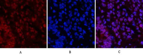
Abbkine has selected the antibody of apoptosis series. After multiple strict verifications, it is suitable for a variety of cell applications and meets the research needs of most customers. The excellent results are as follows: