


Beyond BrdU, New Generation Cell Proliferation Imaging Analysis Kit (EdU Method)
Common methods of cell proliferation mainly include MTT method, WST-1 method and CCK-8 method, which are mainly based on the activity of cells, thus reflecting the overall proliferation effect, but unable to detect individual proliferating cells. Recognized as the most accurate method to detect cell proliferation is to directly detect DNA synthesis in cells. Previously, BrdU method was the most commonly used method. However, the method requires DNA denaturation (such as acid denaturation, thermal denaturation or DNase digestion) to expose BrdU, thus binding to BrdU antibody. The whole experiment has many influencing factors and poor stability.
Abbkine innovates and develops EdU method based on EdU incorporation and subsequent click reaction to solve this application difficulty:
| Product name | Cat# | Fluorescent characteristic |
| Cell Proliferation EdU Image Kit (Green Fluorescence) | KTA2030 | AbFluor 488 Ex/Em = 501/525 nm |
| Cell Proliferation EdU Image Kit (Orange Fluorescence) | KTA2031 | AbFluor 545 Ex/Em = 546/565 nm |
Principle:EdU (5-ethynyl-2´-deoxyuridine) is a nucleoside analog of thymidine and is incorporated into DNA during active DNA synthesis. Comparing to BrdU assays, the EdU-Click Assays are not antibody based and therefore do not require DNA denaturation (typically using HCl or heat or digestion with DNase) for detection of the incorporated nucleoside. Detection is based on a click reaction, a copper-catalyzed covalent reaction between an azide and an alkyne, is complete within 30 minutes.
| Kit components | Advantage |
| • EdU (10mM) • AbFluor 488 azide or AbFluor 545 azide • 10×Reaction buffer • Copper • Reducing Agent | • Not antibody based • No DNA denaturation. Can maintain cell morphology and DNA integrity • Patented AbFluor 488 and 545 azide have good light stability and quenching resistance • Optimization for fluorescence microscope |
Results:
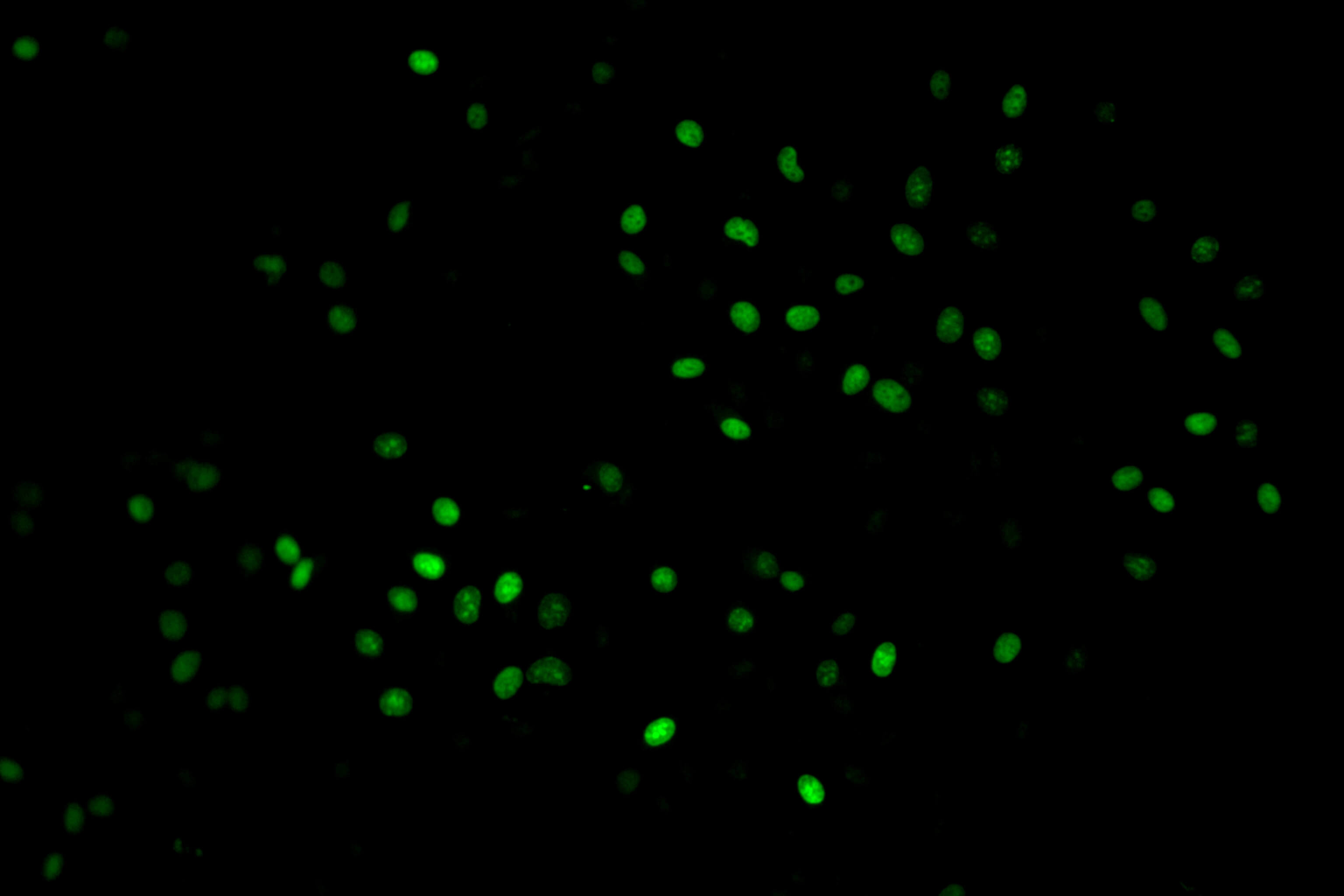 | 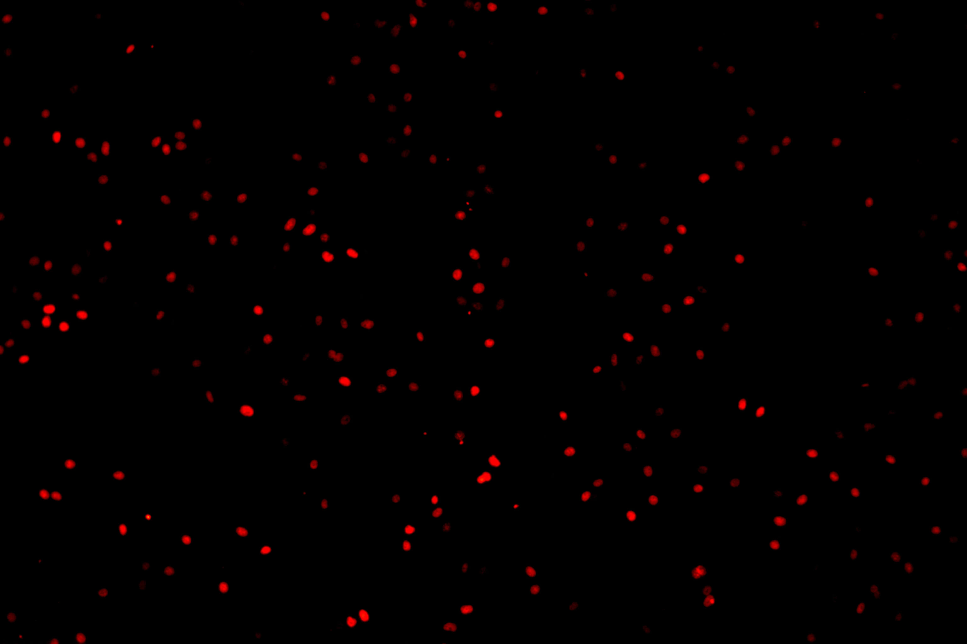 |
| KTA2030 Imaging: Hela Cells (Green) | KTA2031 Imaging: Hela Cells (Red) |


