| Product name | Caspase 9 Monoclonal Antibody |
| Immunogen | Synthetic Peptide |
| Host | Mouse |
| Reactivity | Human,Mouse,Rat,chicken |
| Applications | WB,IHC,IF,IP |
| Applications notes | Optimal working dilutions should be determined experimentally by the investigator. Suggested starting dilutions are as follows: WB 1:1000-5000;IP 1:200;IF 1:200;IHC 1:50-300 |
| Clonality | Monoclonal |
| Preparation method | The antibody was affinity-purified from mouse ascites by affinity-chromatography using specific immunogen. |
| Alternative | CASP9; MCH6; Caspase-9; CASP-9; Apoptotic protease Mch-6; Apoptotic protease-activating factor 3; APAF-3; ICE-like apoptotic protease 6; ICE-LAP6 |
| Formulation | Liquid solution |
| Concentration | 1 mg/ml |
| Molecular weight | 46kD |
| Storage buffer | PBS, pH 7.4, containing 0.5%BSA, 0.02% sodium azide as Preservative and 50% Glycerol. |
| Storage instructions | Stable for one year at -20°C from date of shipment. For maximum recovery of product, centrifuge the original vial after thawing and prior to removing the cap. Aliquot to avoid repeated freezing and thawing. |
| Shipping | Gel pack with blue ice. |
| Precautions | The product listed herein is for research use only and is not intended for use in human or clinical diagnosis. Suggested applications of our products are not recommendations to use our products in violation of any patent or as a license. We cannot be responsible for patent infringements or other violations that may occur with the use of this product. |
| Background | CASP9 encodes a member of the cysteine-aspartic acid protease (caspase) family. Sequential activation of caspases plays a central role in the execution-phase of cell apoptosis. Caspases exist as inactive proenzymes which undergo proteolytic processing at conserved aspartic residues to produce two subunits, large and small, that dimerize to form the active enzyme. Caspase 9 can undergo autoproteolytic processing and activation by the apoptosome, a protein complex of cytochrome c and the apoptotic peptidase activating factor 1; this step is thought to be one of the earliest in the caspase activation cascade. Caspase 9 is thought to play a central role in apoptosis and to be a tumor suppressor. Alternative splicing results in multiple transcript variants. |
| Gene ID | 842 |
| Alternative | CASP9; MCH6; Caspase-9; CASP-9; Apoptotic protease Mch-6; Apoptotic protease-activating factor 3; APAF-3; ICE-like apoptotic protease 6; ICE-LAP6 |
| Others | The antibody detects endogenous Caspase 9 protein. |
| Accession | P55211 |
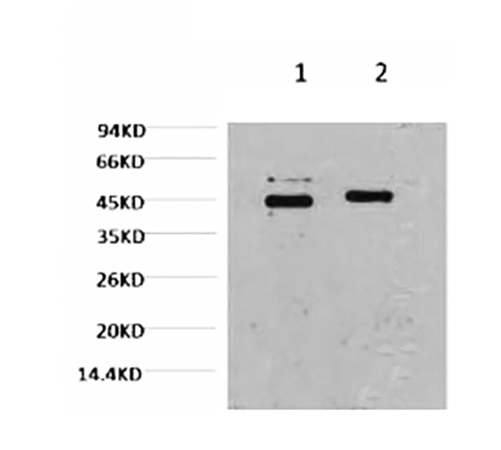
Fig.1. Western blot analysis of Hela, diluted at 1) 1:2000, 2) 1:5000.
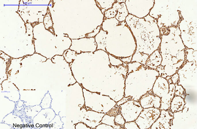
Fig.2. Immunohistochemical analysis of paraffin-embedded human lung tissue. 1, Caspase 9 Monoclonal Antibody was diluted at 1:200 (4°C, overnight). 2, Sodium citrate pH 6.0 was used for antibody retrieval (>98°C, 20min). 3, secondary antibody was diluted at 1:200 (room temperature, 30min). Negative control was used by secondary antibody only.
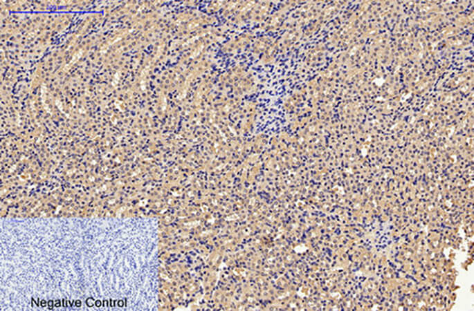
Fig.3. Immunohistochemical analysis of paraffin-embedded mouse kidney tissue. 1, Caspase 9 Monoclonal Antibody was diluted at 1:200 (4°C, overnight). 2, Sodium citrate pH 6.0 was used for antibody retrieval (>98°C, 20min). 3, secondary antibody was diluted at 1:200 (room temperature, 30min). Negative control was used by secondary antibody only.
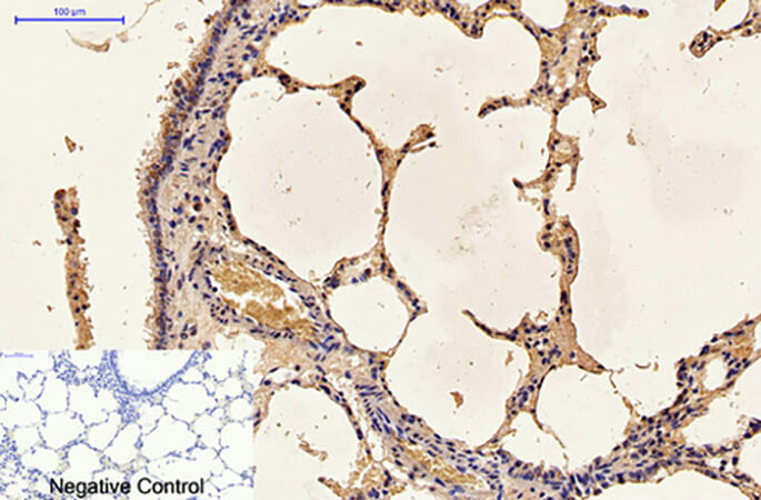
Fig.4. Immunohistochemical analysis of paraffin-embedded rat lung tissue. 1, Caspase 9 Monoclonal Antibody was diluted at 1:200 (4°C, overnight). 2, Sodium citrate pH 6.0 was used for antibody retrieval (>98°C, 20min). 3, secondary antibody was diluted at 1:200 (room temperature, 30min). Negative control was used by secondary antibody only.
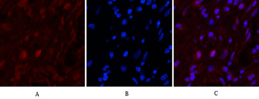
Fig.5. Immunofluorescence analysis of human appendix tissue. 1, Caspase 9 Monoclonal Antibody (red) was diluted at 1:200 (4°C, overnight). 2, Cy3 Labeled secondary antibody was diluted at 1:300 (room temperature, 50min). 3, Picture B: DAPI (blue) 10min. Picture A: Target. Picture B: DAPI. Picture C: merge of A+B.
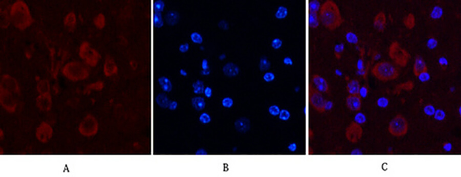
Fig.6. Immunofluorescence analysis of mouse brain tissue. 1, Caspase 9 Monoclonal Antibody (red) was diluted at 1:200 (4°C, overnight). 2, Cy3 Labeled secondary antibody was diluted at 1:300 (room temperature, 50min). 3, Picture B: DAPI (blue) 10min. Picture A: Target. Picture B: DAPI. Picture C: merge of A+B.
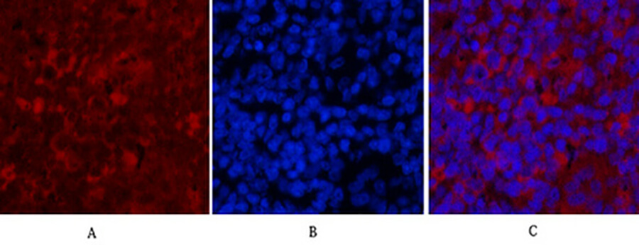
Fig.7. Immunofluorescence analysis of rat spleen tissue. 1, Caspase 9 Monoclonal Antibody (red) was diluted at 1:200 (4°C, overnight). 2, Cy3 Labeled secondary antibody was diluted at 1:300 (room temperature, 50min). 3, Picture B: DAPI (blue) 10min. Picture A: Target. Picture B: DAPI. Picture C: merge of A+B.
Author:Guoqing Hou Publication name:Evid Based Complement Alternat Med IF:5.6
Author:A Liu, X Xu, R Hou, S Badawy, Y Tao, D Chen, A Ihsan Publication name:Toxicology IF:3
You must be logged in to post a review.


Reviews
There are no reviews yet.