


How much we know about cell death
Programmed cell death is an active, orderly and gene-controlled process of cell death, including apoptosis, pyrodeath, autophagy and iron death.
- Apoptosis is the first kind of programmed cell death mode discovered by people, and it is also the most typical and the most clearly studied programmed cell death mode. Apoptosis is the normal physiological response of cells after receiving some specific signal stimulation.
Apoptosis can be divided into two main categories according to its inducible factors: endogenous apoptosis (mitochondrial apoptosis pathway) and exogenous apoptosis (death receptor pathway).
Endogenous apoptosis is mainly controlled by the BCL2 protein family, which includes pro-apoptotic factors BAX, BAK and BOK, and anti-apoptotic factors BCL2, BCLXL, BCLw and MCL1, etc. Through the interaction between pro-apoptotic factors and anti-apoptotic factors, the apoptotic process is regulated jointly.
Exogenous apoptosis is mainly mediated by death receptors such as TNFR1, Fas, DR3 and EDAR on the cell membrane. After the death receptors bind to ligands, they bind to Caspase-2, Caspase-8 and Caspase-10 to form apoptotic complex, thus inducing apoptosis.
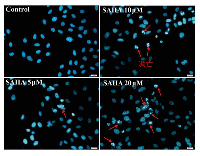
The nuclear structure breaks down in the late apoptotic period, so the observation of nuclear morphology is also a relatively direct indicator. The picture is from the Internet.
- The morphological characteristics of pyroptosis were that pyroptosis cells showed swelling and many bubble-like protrusions under light microscope. Compared with necrotic cells, scorched cells have less swelling. Under electron microscope, it can be clearly seen that the pyrotic cells formed a large number of vesicles, namely pyrotic bodies, before the breakdown of the plasma membrane. Then pores form in the membrane, the membrane bursts, and the contents flow out.
- Autophagy: autophagy was proposed by Ashford and Porter in 1962 after they discovered the phenomenon of "self-eating" in cells. It refers to the formation of autophagosome by the bilayer membrane, which falls off from the ribosome-free region of the rough endoplasmic reticulum and contains degraded organelles, proteins and other components in the cell, and then fuses with lysosomes to form autophagosome, which degrades its contained contents to meet the metabolic needs of the cell itself and renew some organelles. In the process of autophagy, the formation of autophagosome is the key. Its diameter is generally 300~900 nm, with an average of 500 nm. Common inclusions in vesicles include cytoplasmic components and some organelles, such as mitochondria, endocytosomes, and peroxisome. Compared with other organelles, the half-life of autophagy is very short, only about 8 minutes, indicating that autophagy is an effective response of cells to environmental changes.
- Iron death: Iron death is an iron-dependent and peroxide-driven form of cell death associated with a variety of metabolic disorders and homeostasis. The Fenton reaction between iron ions and unsaturated fatty acids on the cell membrane produces a large number of reactive oxygen species. When the antioxidant capacity of the cell is exceeded, oxidative stress reaction occurs, which damages mitochondria, endoplasmic reticulum and nucleic acid in the cell, thus leading to cell death. The expression of antioxidant system (glutathione GSH and glutathione peroxidase 4-GPX4) was also decreased. The main morphological characteristics of iron death cells: Iron death can lead to the reduction of mitochondria, membrane density, ridge reduction. The morphological changes in the nucleus were not obvious.
5, Copper death: Copper death (Cuproptosis) is a newly discovered cell death phenomenon, that is, the imbalance of copper ion homeostasis induced cell death process. Copper death occurs through direct combination of copper with the esterified component of the tricarboxylic acid cycle. This leads to acylated protein aggregation and subsequent loss of ferrithiolin, which causes toxic stress of the protein and ultimately leads to cell death.
The research on cell death is extensive and profound. Under his unremitting efforts, Iacoin has conducted in-depth research in the direction of apoptosis and developed a series of apoptosis detection kits, which mainly include the following products:
| Product NO. | Product name | Size |
| KTA0005
|
Annexin V-EGFP/PI Apoptosis Detection kit | 20T/50T/100T |
| KTA0001
|
Annexin V-AbFluor™ 405 Apoptosis Detection kit (Blue Fluorescence) | 50T/100T |
| KTA0002
|
Annexin V-AbFluor™ 488 Apoptosis Detection kit (Green Fluorescence) | 50T/100T |
| KTA0003
|
Annexin V-AbFluor™ 555 Apoptosis Detection kit (Orange Fluorescence) | 50T/100T |
| KTA0004
|
Annexin V-AbFluor™ 647 Apoptosis Detection kit (Red Fluorescence) | 50T/100T |
| KTA4001 | Mitochondrial Membrane Potential Assay Kit (JC-1) | 20T/100T/500T |
| KTA3020 | Caspase-1 Assay Kit (Colorimetric) | 20T/50T/100T |
| KTA3021 | Caspase-2 Assay Kit (Colorimetric) | 20T/50T/100T |
| · KTA2010 | One-step TUNEL Apoptosis Assay Kit (Green Fluorescence) | 50T/100T |
| · KTA2011 | One-step TUNEL Apoptosis Assay Kit (Orange Fluorescence) | 50T/100T |
Annexin V-AbFluor™ 488 Apoptosis Detection kit (Green Fluorescence) application:
1.Microscope double-dye photography
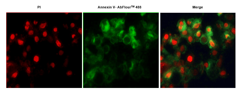
Fig.1. Hela cells were induced with camptothecin for 24 hours and stained with Annexin V- AbFluor™ 488/PI Apoptosis Detection Kit. The cell is a late stage apoptotic/necrotic cell with both Annexin V- AbFluor™ 488 and PI staining (green membrane with red fragmented nucleus).
2.Apoptosis analysis by flow cytometry:

Fig.2. Hela cells were induced with camptothecin for 24 hours and stained with Annexin V- AbFluor™ 488 Apoptosis Detection Kit. The combination of AbFluor™ 488 and propidium iodide allows for the distinction between early apoptotic cells (Annexin V- AbFluor™ 488 positive), late apoptotic and/or necrotic cells (Annexin V- AbFluor™ 488 and propidium iodide positive), and viable cells (unstained).
Abbkine focuses on the field of proteomics and cytology, focusing on the innovation and development of various types of antibodies, proteins, analytical reagents and kits, with a view to becoming a key enabler in the field of life science research and development, drug research and development. We offer your favorite products for protein and immune research, from basic immunological products such as protein extraction and quantification, to internal reference tag antibodies, primary and secondary antibodies for immunological experiments, etc. Cell research users' favorite products, from dyes and kits for cell status detection, organelle extraction kits, cell substructure staining tracer and cell metabolism detection products, to cytokine and protein detection kits for cell culture, just to help your research career!


