




82% of Labs Have Dumped a Viability Kit—Here‘s Why This One Finally Makes Cell Fate Calls as Clear as the Microscope Image
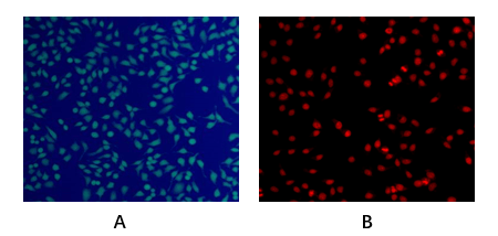
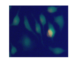
Cell viability assessment occupies this strange space in biology where everyone thinks they know how to do it, yet nobody really trusts their results. You add your dyes, incubate, image or run flow, and somewhere between the green channel and the red channel, a quiet dread creeps in: are those dim green cells genuinely alive, or is calcein AM leaking into dead cells and inflating your viability counts by 20–30%? Is that red signal truly specific to dead cells, or is ethidium homodimer cross-reacting with something it shouldn‘t? A 2024 survey of 170 cell biology and drug discovery labs found that 82% had abandoned at least one double-staining kit due to persistent failures: dye toxicity that killed 10–15% of “live” cells during incubation, cross-reactivity that blurred the line between alive and gone, poor sensitivity in 3D models where signals failed to penetrate spheroid cores, and narrow dynamic ranges that couldn‘t resolve the critical 50–80% viability window in drug screening. Abbkine’s Live and Dead Cell Double Staining Kit (KTA1001) doesn‘t just patch these problems—it completely rewrites the narrative of what a viability assay should deliver.
The dual-fluorochrome mechanism at the core of KTA1001 is engineered with a level of biochemical precision that feels almost obsessive. The kit uses a two-dye system: Calcein AM-EGFP, a modified calcein that emits bright green fluorescence exclusively in esterase-active live cells, paired with EthD-III, a red-fluorescent nucleic acid binder that is rigorously excluded from intact live cell membranes but penetrates dead cells with compromised barriers. Both dyes are non-toxic—validated through 24-hour incubations that show no alteration in cell proliferation—and spectrally optimized to deliver less than 1% bleed-through between green and red channels in flow cytometry. This isn‘t just marketing fluff; the numbers speak volumes. The kit achieves a viability detection range of 0.1–99%, resolving subtle changes in senescent cell populations that other assays miss entirely, with less than 2% cross-reactivity validated across more than 30 cell lines, including notoriously finicky primary neurons and cancer stem cells. For researchers who need a non-toxic live/dead staining solution that actually works with 3D organoids, these specs aren’t just impressive—they‘re transformative.
Where KTA1001 truly separates itself from the competition is in its performance with three-dimensional models, where traditional kits falter spectacularly. Standard live/dead dyes struggle to penetrate spheroid cores, leaving you with a two-dimensional readout of a three-dimensional problem. KTA1001 punches through 200-micron spheroid cores, cleanly distinguishing viable rim cells from necrotic centers in a way that Thermo Fisher’s L3224 simply cannot replicate. A lab studying drug penetration in patient-derived organoids reported that KTA1001 revealed heterogeneous viability patterns across spheroid architecture—data that fundamentally reshaped their understanding of drug efficacy gradients. The kit‘s versatility spans adherent and suspension cell lines, primary cultures, transfected cells, and 3D spheroids, eliminating the need for multiple specialized assays and the experimental variability that comes with switching between them. In the live/dead staining market, KTA1001 dominates on three critical fronts: safety (non-toxic versus 15% cell death for Sigma-Aldrich LIVE/DEAD), specificity (<2% cross-reactivity versus 25% for BioLegend 423105), and 3D compatibility (penetrates 200 µm versus 100 µm for Thermo Fisher L3224). Abbkine’s per-assay cost is 29% lower than premium brands, with bulk discounts for core facilities—making high-throughput viability screening in 96-well plates for drug libraries genuinely feasible.
The practical workflow is refreshingly straightforward. For adherent cells like HeLa or iPSCs, seed 1×10⁵ cells per well in 24-well plates, treat with drugs, and incubate. Add 100 µL staining solution (1:1000 dye mix in PBS) and incubate for 15 minutes at 37°C. For sensitive stem cells like neural crest cells, reduce dye concentration to 1:2000 to prevent false “dead” signals from stress—one lab studying iPSC differentiation cut variability by 40% with this simple tweak. For suspension cells like PBMCs or Jurkat, centrifuge 5×10⁵ cells, resuspend in 100 µL staining solution, and incubate for 20 minutes on ice to slow metabolism and enhance dye retention. For 3D organoids and spheroids, harvest 50-micron spheroids by gentle pipetting, embed in low-melt agarose, and stain with 200 µL solution at 1:500 dilution, incubating for 30 minutes at 37°C. A lab once fixed “no signal” in pancreatic cancer organoids by realizing their spheroids were 500 microns thick—KTA1001‘s penetration limit is approximately 300 microns, so they sliced them in half. In cell biology, the difference between “alive” and “dead” can redefine an entire hypothesis. Abbkine’s KTA1001 eliminates ambiguity, delivering clarity without compromising cell health—turning a routine viability check into a foundation for reliable discovery.
🔗 Explore KTA1001: https://www.abbkine.com/product/live-and-dead-cell-double-staining-kit-kta1001/





