




The Kitchen Secret That Cushions a Nectarine—And the Enzyme That Quietly Destroys a $3.2 Billion Cold Chain
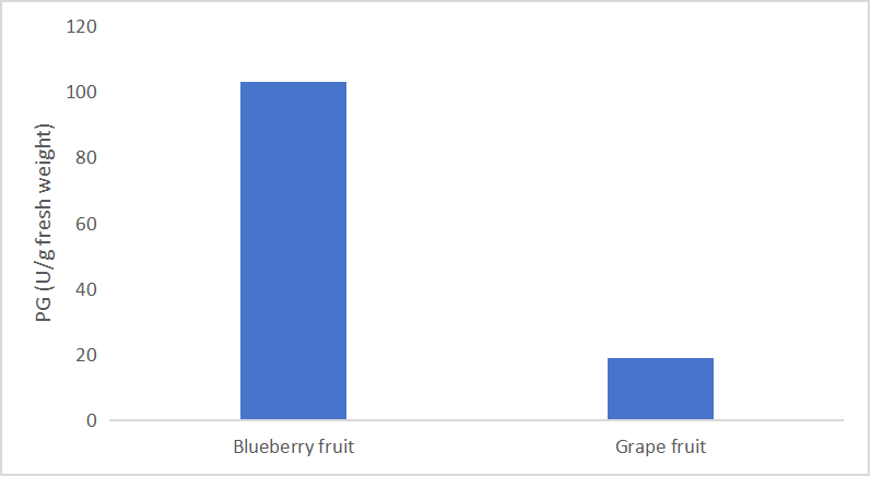
If you have ever watched a perfect, unbruised strawberry dissolve into a sunken pool of juice and gray mold exactly thirty‑six hours after you brought it home from the market, you have witnessed polygalacturonase in action. The enzyme does not announce itself. It does not require oxygen, light, or the cooperation of the fruit’s still‑living cells. It is secreted into the apoplast—the labyrinthine space between plant cell walls—where it hydrolyzes the α‑1,4‑glycosidic bonds that hold the galacturonic‑acid backbone of pectin together, and the middle lamella, the adhesive cement that glues one cell to the next, progressively dissolves. What we call “softening” is, at the molecular level, polygalacturonase‑mediated solubilization of cell‑wall pectin. What we call “spoilage” is, in many cases, the same enzymatic cascade commandeered by Botrytis cinerea or Penicillium expansum to breach the host cell wall before the innate immune system can mount a defense. Polygalacturonase (PG, EC 3.2.1.15) is widely distributed across plants, bacteria, and fungi, and its biological footprint extends into processes as diverse as fruit ripening and abscission, pollen tube emergence, seed maturation, and pathogen virulence. It is, simultaneously, a commercial liability measured in the billions of dollars and one of the most biochemically significant hydrolases in the plant kingdom. A 2018 review in Postharvest Biology and Technology noted that excessive softening of fruit is a major cause of post‑harvest losses throughout the world, contributing to wastage of up to half of the harvested crop. The enzyme that drives that loss has, for decades, been easier to talk about than to measure. Researchers have characterized PG at the transcript level, quantified its protein abundance by western blot, and inferred its activity from changes in fruit firmness or pectin molecular weight distributions, but the defining catalytic parameter—the rate of polygalacturonic‑acid hydrolysis—has remained, for many laboratories, inaccessible.
That inaccessibility is not for lack of a workable assay. The 3,5‑dinitrosalicylic acid (DNS) method has been the gold standard for glycoside hydrolase activity measurement since it was first adapted for reducing‑sugar detection in the middle of the twentieth century. PG hydrolyzes polygalacturonic acid to liberate galacturonate residues, each of which carries a free reducing aldehyde group that reduces DNS to 3‑amino‑5‑nitrosalicylic acid under alkaline conditions at high temperature, forming a reddish‑brown chromophore with a characteristic absorption at 540 nm. The chemistry is robust, the stoichiometry is well characterized, and the readout is visible on any standard spectrophotometer. The problem has never been the DNS reaction. The problem has been the manual execution: separate boiling steps for each sample, fume‑hood handling of hot alkaline reagents, per‑sample cuvette readings that make population‑level phenotyping logistically punitive, and the universal interference from endogenous reducing sugars and non‑PG pectinolytic enzymes that co‑purify with the target activity and generate background signals indistinguishable from genuine PG output. For a postharvest biologist who needs to screen 200 fruit genotypes across four ripening stages, manual DNS simply does not scale. For a plant pathologist quantifying PG secretion from Erwinia carotovora cultures across a treatment matrix, the throughput gap between the biochemical gold standard and the experimental demand has forced the field to rely on surrogates—transcript abundance, immunoassay signal, or simple penetrometer measurements—that are informative but are not PG activity.
Abbkine’s CheKine™ Micro Polygalacturonase (PG) Activity Assay Kit (KTB1333) relocates the DNS‑based detection chemistry into a microplate format that multiplies the practical throughput of PG measurement without abandoning the biochemical principle that has made the DNS method an analytical mainstay for over seventy years. PG in the sample hydrolyzes the provided polygalacturonic acid substrate to release galacturonate residues bearing reducing aldehyde groups; those residues react with the DNS reagent included in the kit under alkaline heating conditions to produce a stable reddish‑brown reaction product whose absorbance at 540 nm is directly proportional to PG activity. There are no secondary enzymatic cascades, no antibody‑based detection steps, no requirement for a fluorescence detector, and no need for the radioisotope‑labeled pectin substrates that a previous generation of PG enzymologists was forced to synthesize in‑house. The signal reaching the plate reader is the same DNS chromophore that has been the workhorse of carbohydrate enzymology since the 1950s, now generated by pre‑formulated, stabilized reagents optimized for a 96‑well plate and a visible‑wavelength plate reader.
The operational difference between manual DNS and KTB1333’s microplate implementation is not incremental; it is qualitative. Manual DNS protocols require individual sample tubes, individual boiling‑water‑bath steps, and individual spectrophotometer readings that force the operator to track absorbance values for each sample against a stopwatch. Variability in heating time, cooling time, and pipetting cadence accumulates across samples until the resulting dataset contains as much protocol‑induced variation as biological variation. KTB1333 consolidates the DNS reaction into a streamlined microplate workflow where samples, blanks, and standards are processed in parallel under identical thermal conditions, the absorbance is read across the entire plate in seconds, and the rate of color development reflects the PG activity in each well rather than the operator’s pipetting speed. The practical implications for throughput are immediate: a dose‑response matrix spanning five elicitor concentrations, four time points, and three biological replicates—sixty samples—can be assayed in a single 96‑well plate in under an hour.
The sample compatibility statement is comprehensive: plant tissues (the primary application domain), bacteria, fungi, and liquid samples. The inclusion of bacterial and fungal samples alongside plant tissue is not a marketing afterthought; it acknowledges that PG is equally relevant as a plant developmental enzyme and as a pathogen‑secreted virulence factor. When Botrytis cinerea penetrates a ripening strawberry achene, it secretes a cocktail of cell‑wall‑degrading enzymes in which PG is among the earliest and most biochemically potent, and quantifying PG activity in fungal culture filtrates is a direct measurement of a pathogenicity determinant. When Erwinia carotovora causes soft‑rot disease in potato tubers, the maceration of host tissue is largely attributable to pectinolytic enzyme activity, and PG is a primary contributor. The kit’s stated compatibility with bacteria and fungi opens the door to PG activity measurement in experimental systems that span plant developmental biology, postharvest physiology, plant‑pathogen interactions, and microbial biotechnology.
The component architecture is deliberately compact: Extraction Buffer, Reagent I, Reagent II, and a Standard. No separate DNS reagent preparation that must be performed in a fume hood. No polygalacturonic acid substrate that must be sourced from a specialty chemical supplier and titrated for each new lot. No boiling‑water‑bath step that requires open‑flame or heating‑block access and generates fumes incompatible with a standard laboratory bench. Storage is at 4 °C, protected from light, with a six‑month stability window from receipt. Shipping is on gel packs with blue ice. Fresh samples are recommended for optimal results, and the protocol advises that if the assay cannot be performed immediately, completing the sample‑preparation step before storing at ‑80 °C preserves enzyme activity for up to one month. The standard laboratory discipline of performing several dilutions to confirm readings fall within the standard value range is stated plainly—an instruction that reflects genuine analytical practice rather than a generic disclaimer.
The product page currently shows zero publications citing KTB1333. Zero citations for a recently launched enzyme activity kit targeting an enzyme whose measurement has been constrained to manual protocols is not a red flag. It reflects the launch timing of the product. What matters is the design: DNS‑based detection chemistry adapted for a microplate format, substrate‑specific detection of PG‑generated reducing ends, a sample compatibility list spanning plant tissues, bacteria, fungi, and liquid samples, and a component architecture that eliminates the reagent‑preparation burden that has historically restricted manual PG assays to laboratories with dedicated carbohydrate‑chemistry expertise. These are the specifications that generate citations, not the other way around.
The broader biological context makes a strong case for routine PG activity measurement that extends well beyond the postharvest‑biology applications for which PG is most frequently cited. In plant‑pathogen interactions, PG is one of the earliest secreted virulence factors, and its activity is directly targeted by host‑encoded polygalacturonase‑inhibiting proteins (PGIPs) that bind PG and suppress cell‑wall degradation while simultaneously generating oligogalacturonide fragments that activate pattern‑triggered immunity. A 2024 study published in Science demonstrated that PGIPs hijack pathogen‑derived polygalacturonase activity to produce immunity‑eliciting oligogalacturonides, converting a virulence factor into a trigger for defense. In that experimental context, PG activity is not a biomarker of disease; it is the biochemical interface where pathogen offense and host defense physically collide, and the ability to measure PG activity quantitatively across pathogen strains, host genotypes, and PGIP concentrations is essential to dissecting the interaction. In fruit development, antisense down‑regulation of the polygalacturonase gene FaPG1 in strawberry produced fruit that remained firm significantly longer than wild‑type controls, providing direct genetic evidence that PG activity is the primary determinant of softening in this commercially critical crop. For the fruit breeder screening a germplasm collection for firmness retention, PG activity is a phenotypic parameter as informative as soluble solids content or titratable acidity, and its measurement at scale has been logistically prohibitive until the advent of microplate‑adapted colorimetric detection.
For the postharvest physiologist screening storage treatments for firmness retention in stone fruit, the plant pathologist quantifying PG secretion as a virulence phenotype, the fruit geneticist mapping QTL that determine shelf life, the microbial biotechnologist engineering fungal strains for industrial pectinase production, or the plant biologist characterizing cell‑wall remodeling during organ abscission and pollen tube emergence, direct PG activity measurement converts the observation of softening into a quantitative biochemical dataset. The kitchen secret that cushions a nectarine and the enzyme that destroys a cold‑chain shipment are the same molecule. KTB1333 makes that molecule measurable, directly and at scale.
Explore specifications, access the protocol, and place your order here: https://www.abbkine.com/product/chekine-mirco-ploygalacturonase-pg-activity-assay-kit-ktb1333/





