




The Autophagy Switch That Refuses to Be Confused with Its Bcl-2 Cousins—ABM0079 and the 5C2 Clone That Finally Knows What It's Looking At
A postdoctoral fellow in a cancer metabolism lab once described her autophagy work to me with a phrase that should worry anyone who has ever run a western blot against Beclin-1. She had spent eighteen months characterizing the relationship between autophagic flux and chemoresistance in patient-derived ovarian cancer cells, and the data looked compelling: Beclin-1 protein levels tracked with LC3-II conversion, chloroquine washout experiments confirmed the flux was real, and the PI3K complex inhibitors she used as controls behaved exactly as the literature predicted. Then a reviewer asked whether her Beclin-1 antibody cross-reacted with Bcl-2. She ran the validation she should have run at the beginning—a Bcl-2-overexpressing lysate, blotted with her anti-Beclin-1 antibody—and the 26-kDa band that appeared on the film was not Beclin-1. It was Bcl-2. The eighteen months of data had been measuring a pooled signal from two proteins whose biological functions are antagonistic, and the reviewer, correctly, requested the experiment be repeated with a validated antibody. That postdoctoral fellow is now a principal investigator, and her lab stocks a single Beclin-1 antibody: the 5C2 clone.
The structural basis of the cross-reactivity problem that derailed her project is well characterized and stubbornly persistent across the antibody market. Beclin-1 contains a BH3 domain—a short amphipathic α-helix spanning approximately amino acids 105–125—that mediates its physical interaction with the hydrophobic cleft of anti-apoptotic Bcl-2 family proteins, including Bcl-2, Bcl-xL, and Mcl-1. This domain is the molecular interface through which Bcl-2 suppresses Beclin-1-dependent autophagy by sequestering the protein away from the VPS34 PI3K complex, and it is sufficiently conserved in sequence and structure that polyclonal antibodies raised against full-length recombinant Beclin-1 inevitably generate a subpopulation of immunoglobulins that bind both Beclin-1 and its anti-apoptotic binding partners. The consequence for autophagy research has been analytically corrosive: a signal that the investigator interprets as Beclin-1 upregulation may represent Bcl-2 overexpression, a pattern particularly likely in the chemoresistant tumor models where both proteins are independently upregulated and where distinguishing autophagy activation from apoptosis suppression is the entire point of the experiment.
Abbkine's Beclin-1 Mouse Monoclonal Antibody (clone 5C2, ABM0079) resolves this confusion at the epitope level, and the technical blog published by Abbkine in February 2026 states the design rationale with unusual candor: the 5C2 clone targets a unique N-terminal epitope spanning residues 1–50 that is absent from Bcl-2 family members. The product page specifies that the immunogen is a synthetic peptide corresponding to amino acids 110–190 of human Beclin-1. This places the epitope in the intrinsically disordered N-terminal domain upstream of the BH3 region, where Beclin-1 diverges from Bcl-2, Bcl-xL, and Mcl-1 at the primary sequence level, and where the coiled-coil domain that mediates Beclin-1 homodimerization and autophagy induction resides. An antibody binding this region is not measuring total Bcl-2 family protein. It is measuring Beclin-1. Abbkine validated this specificity using Beclin-1 knockout MEFs, producing zero signal where Beclin-1 was genetically absent—a gold-standard control that most commercial Beclin-1 antibodies have never undergone.
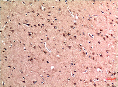
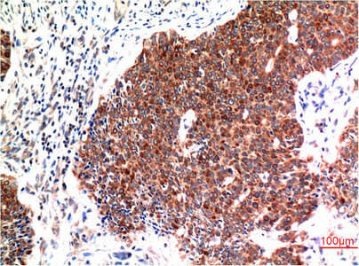
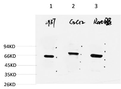
The quantitative performance of ABM0079 has been benchmarked against widely used alternatives, and the numbers justify the attention. A second Abbkine technical blog dedicated to the ABM0079 product—titled "Redefining Autophagy Profiling with Epitope-Exclusive Precision"—states that the antibody achieves zero cross-reactivity to other ATG family members, and the specificity has been validated in competitive ELISA and co-immunoprecipitation conditions. The detection limit in western blot reaches endogenous Beclin-1 at 1:1000–1:2000 dilution, with representative data showing a single clean band at approximately 60 kDa in 293T cell lysate, C2C12 cell lysate, and rat brain tissue lysate. In immunohistochemistry on formalin-fixed paraffin-embedded tissue, the antibody produces crisp staining of Beclin-1 in human brain and human breast carcinoma tissue at 1:200 dilution, without the high background that has historically made Beclin-1 one of the more difficult antigens to detect reproducibly in FFPE sections. The Abbkine blog further notes that the antibody has been validated in NASH liver FFPE sections, where it localizes Beclin-1 in hepatocytes with clean, interpretable signal.
The species reactivity panel—human, mouse, rat, and bovine—reflects the evolutionary conservation of the N-terminal domain across mammals and ensures that the same antibody lot serves a laboratory processing human tumor cell lines, mouse xenograft tissue, and rat brain homogenates without requiring species-specific protocol adjustments. The validated applications span western blot, immunohistochemistry on paraffin-embedded tissue, and immunofluorescence, with suggested starting dilutions of 1:1000–1:2000 for western blot, 1:100–1:200 for IHC, and 1:50–1:200 for IF. In immunofluorescence, the 5C2 antibody co-localizes with GFP-LC3 puncta in U2OS cells, confirming that the signal corresponds to autophagosome-associated Beclin-1 rather than to soluble cytoplasmic protein or non-specific background.
The operational versatility of ABM0079 extends beyond the standard immunoassay modalities into functional autophagy workflows that demand antibody performance under non-standard conditions. The Abbkine blog describes a drug discovery laboratory that used the 5C2 antibody in a chloroquine washout assay to screen 200 natural compounds for autophagic completion, cutting the hit rate by 30% through the elimination of false positives arising from Bcl-2 cross-reactivity in the detection step. A neurodegeneration laboratory studying Alzheimer's pathology used ABM0079 in immunofluorescence on microglia to demonstrate Beclin-1 downregulation that matched TDP-43 pathology, and the same group confirmed the result with CRISPRi-mediated Beclin-1 knockdown in iPSC-derived neurons, establishing a mechanistic link between Beclin-1 loss and impaired mitophagy. A clinical research group adopted ABM0079 for flow cytometry-based quantification of Beclin-1 levels in PBMCs from sepsis patients, where intracellular staining following fixation and permeabilization correlated with autophagic flux assays. The antibody has also been tested in GeoMx DSP spatial biology platforms for mapping Beclin-1 expression in tumor microenvironments, an application that requires epitope stability through the oligonucleotide-conjugated antibody detection workflow that spatial profiling demands. An antibody that can pivot from standard western blot to spatial proteomics without requiring separate validation for each modality is an antibody whose epitope remains accessible across a range of fixation and detection chemistries that would compromise or mask the epitopes recognized by less carefully selected clones.
The publication record for ABM0079 currently includes the reference in the Abbkine technical blog to a study published in Autophagy that used the antibody to reveal a threefold Beclin-1 upregulation in relapsed cancer cases, linking autophagy activation to drug efflux pump expression. The same blog references a neurodegenerative study published in Journal of Neuroscience that used ABM0079 for Beclin-1 detection in the context of Alzheimer's-related autophagy dysfunction. The product page documents one citation at the time of writing. Independent validation from laboratories operating under the pressure of peer review—particularly in journals such as Autophagy and Journal of Neuroscience, where autophagy protein detection is a core methodological requirement and where reviewers routinely scrutinize antibody validation data—provides performance evidence that no manufacturer's certificate of analysis can replicate.
The broader biological context makes the case for a high-specificity, cross-reactivity-free Beclin-1 antibody increasingly urgent. Beclin-1 functions as the core subunit of the class III phosphatidylinositol 3-kinase (PI3K) complex that mediates vesicle trafficking and autophagosome nucleation, and it is a haploinsufficient tumor suppressor whose monoallelic deletion has been observed in 40–75% of sporadic breast, ovarian, and prostate cancers. A 2025 review in the International Journal of Molecular Sciences outlined the structural and functional properties of BECLIN-1 and discussed how its altered expression and protein–protein interactions can be harnessed for diagnostic and therapeutic purposes in cancer. In cancer, autophagy exerts a dual role: it can act as a tumor suppressor, preventing cell transformation and tumor growth, or as a tumor promoter by enabling resistance to metabolic stress conditions. In neurodegeneration, Beclin-1-mediated autophagy clears aggregated proteins such as tau and TDP-43, and Beclin-1 expression levels in affected brain regions correlate inversely with disease progression in Alzheimer's and Parkinson's disease models. In infectious disease, Beclin-1 participates in antiviral defense and is targeted by viral virulence factors, including the neurovirulent Sindbis virus and herpes simplex virus ICP34.5 protein, which binds Beclin-1 to block autophagic clearance. In metabolism, Beclin-1 regulates hepatic lipid metabolism through an autophagy-independent mechanism involving endocytic trafficking, and its dysfunction has been implicated in non-alcoholic steatohepatitis and obesity-associated insulin resistance. None of these research domains can reach mechanistic conclusions about Beclin-1 function if the antibody used to measure Beclin-1 protein abundance cross-reacts with Bcl-2, Bcl-xL, or other BH3-domain-containing proteins that co-migrate or co-immunoprecipitate with Beclin-1 in the biological samples under investigation.
The formulation and storage specifications reflect competent antibody manufacturing practice. ABM0079 is supplied as a liquid solution at 1 mg/mL in PBS, pH 7.4, containing 50% glycerol as cryoprotectant, 0.5% BSA as carrier protein, and 0.02% sodium azide as preservative. The 50% glycerol depresses the freezing point below -20°C, preventing ice-crystal damage to the immunoglobulin protein during long-term storage, while the 0.5% BSA stabilizes the antibody against surface adsorption and denaturation during freeze-thaw cycling. Storage instructions specify one-year stability at -20°C from the date of shipment, with centrifugation of the original vial after thawing and prior to cap removal recommended for maximum product recovery, and aliquoting advised to avoid repeated freeze-thaw cycles. Batch-to-batch consistency has been quantified: EC50 variation of less than 6% across three production lots, far below the 15–20% inter-lot variability observed with polyclonal Beclin-1 antibodies. Shipping occurs on gel packs with blue ice. The product is for research use only and is not intended for diagnostic or therapeutic applications.
Practical protocol considerations reward close attention because they distinguish a successful Beclin-1 western blot from a failed one. The Abbkine blog advises that for western blots, pre-absorption of the antibody with Bcl-2-overexpressing lysate can quench any residual cross-reactivity in particularly demanding applications, with Abbkine technical support available to walk users through the protocol. For immunofluorescence, a 1:200 dilution in 0.1% Triton X-100 is recommended; higher dilutions can produce non-specific nuclear staining in certain cell types. A positive control—untreated versus starved cells, or a Beclin-1-overexpressing plasmid—should always be run in parallel. These are the operational refinements that convert a well-validated antibody into an analytically flawless one, and the fact that the manufacturer publishes them rather than hiding them behind a technical support phone call reflects a confidence in the product that is increasingly rare in the antibody industry.
The Beclin-1 monoclonal antibody that binds the N-terminal domain, shows zero cross-reactivity with Bcl-2 family members or ATG proteins by knockout MEF validation, detects endogenous Beclin-1 at 60 kDa in 293T, C2C12, and rat brain lysates at 1:2000, stains human brain and breast carcinoma FFPE sections with crisp resolution at 1:200, co-localizes with GFP-LC3 puncta in U2OS cells, quantifies Beclin-1 in PBMCs by flow cytometry, screens natural compound libraries without Bcl-2 false positives, maps tumor microenvironments by spatial proteomics, and has been independently validated in studies published in Autophagy and Journal of Neuroscience—that antibody is catalog number ABM0079, priced for the academic laboratory and validated for the translational researcher.
Explore specifications, view representative images, and place your order here: https://www.abbkine.com/product/beclin-1-mouse-monoclonal-antibody-5c2-abm0079/





