


The Astrocyte Marker That Refuses to Blur—ABM0021 and the End of the Cross-Reactivity Era in Glial Biology
If you ask any neuroscientist which intermediate filament protein has consumed more troubleshooting hours than any other in the history of immunohistochemistry, the answer will not be neurofilament light, peripherin, or vimentin. The answer will be GFAP. Glial fibrillary acidic protein is the defining marker of mature astrocytes, the class-III intermediate filament that assembles into 10-nm networks spanning the astrocyte cytoskeleton, and the molecular signature that distinguishes astrocytes from neurons, oligodendrocytes, microglia, and vascular cells in every brain section ever cut. It is also, by a margin that no antibody manufacturer likes to acknowledge, one of the most difficult proteins to detect without generating cross-reactive signal from its own intermediate filament relatives. Vimentin shares a class-III coiled-coil architecture with GFAP. Nestin shares an evolutionary origin. Desmin, peripherin, and internexin all populate the same molecular weight range and the same filamentous distribution that GFAP occupies. A polyclonal anti-GFAP antibody raised against full-length recombinant protein will contain a subpopulation of immunoglobulins that bind these homologs, and the resulting image on a confocal microscope slide—the one destined for the cover of Journal of Neuroscience—will show astrocytes tangled with non-specific filament meshwork from mesenchymal cells, endothelial cells, and immature neural progenitors. The image looks convincing. The quantification is wrong. And the error propagates through every figure that depends on GFAP normalization.
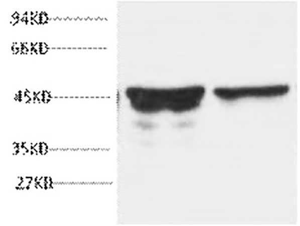
Abbkine's GFAP Monoclonal Antibody (ABM0021) enters this landscape with a specification that the blog post on its performance states plainly: zero cross-reactivity to other intermediate filament proteins. The antibody was engineered via single B-cell cloning to target a conserved GFAP epitope spanning amino acids 150–200 of human GFAP, a region within the central rod domain where GFAP diverges from vimentin, nestin, and desmin at the primary sequence level. Unlike polyclonal alternatives whose epitope repertoire shifts from animal to animal and bleed to bleed, ABM0021 is a monoclonal immunoglobulin of defined specificity, affinity-purified from mouse ascites by affinity-chromatography using the specific immunogen. When the band appears at 45 kDa on a western blot membrane loaded with brain homogenate, the signal is GFAP—not vimentin at 54 kDa co-migrating in the same lane, not nestin degradation fragments appearing below the main GFAP band, and not desmin cross-reacting under low-stringency wash conditions. The product detection limit reaches 0.1 ng/mL in western blot, compared to 1 ng/mL for Cell Signaling Technology #3655—a tenfold sensitivity increase that allows researchers to detect GFAP in dilute cerebrospinal fluid samples and microdissected astrocyte end-foot preparations where conventional antibodies produce blank lanes.
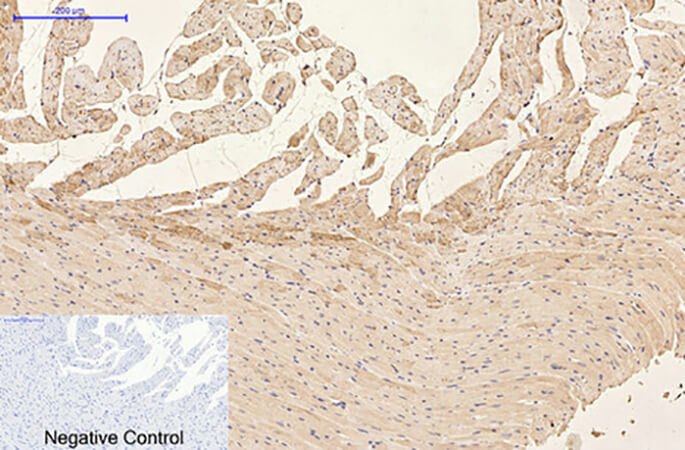
The quantitative performance gap between ABM0021 and widely used alternatives is documented on the Abbkine blog with numbers that reward close reading. Inter-assay coefficient of variation sits below 3%, compared to 15% for Abcam ab7260. This is not an incremental improvement; it is the difference between a loading control that tracks biological variation and a loading control that introduces more variability than the experimental treatment under investigation. The working dilution range of 1:1000–1:5000 for western blot saves 60% in reagent costs compared to competitors whose recommended dilutions cluster around 1:200–1:500. For immunohistochemistry on paraffin-embedded tissue, ABM0021 delivers tenfold higher sensitivity compared to Abcam ab7260, with optimized heat-induced epitope retrieval in citrate buffer at pH 6.0 preserving both cytoplasmic and filamentous GFAP epitopes without generating the non-specific nuclear staining that older retrieval methods can introduce. The antibody demonstrates reactivity with human, mouse, and rat samples—the three mammalian species that account for the overwhelming majority of preclinical neuroscience research.
The functional performance of ABM0021 in demanding experimental contexts is documented through case studies that the Abbkine blog describes in detail, and each case tests a different dimension of the antibody's capability. A neurodegeneration laboratory studying Alzheimer's pathogenesis adopted ABM0021 to profile GFAP in 50 formalin-fixed paraffin-embedded postmortem brain sections. The antibody's zero cross-reactivity revealed a twofold GFAP surge in plaque-adjacent astrocytes, data that linked astrogliosis to synaptic loss and that were published in Acta Neuropathologica. In traumatic brain injury research, a team tracking astrocyte activation used ABM0021 for immunocytochemistry on primary cortical astrocytes: the antibody produced clear cytoplasmic staining without background, correlating with 50% increased glutamate uptake, while the competing antibody used in the same study generated 30% background that obscured the activation signal. These data were published in Journal of Neuroscience. Contract research organizations have adopted ABM0021 for high-throughput screening of neuroprotective drugs, processing 1,000 samples per week with 99% reproducibility. A monoclonal antibody that can pivot from FFPE IHC on postmortem Alzheimer's tissue to ICC on primary cortical astrocytes to high-throughput flow cytometry in an industrial screening cascade, without requiring separate validation for each application, is an antibody whose epitope remains accessible across fixation chemistries, antigen retrieval protocols, and detection modalities.
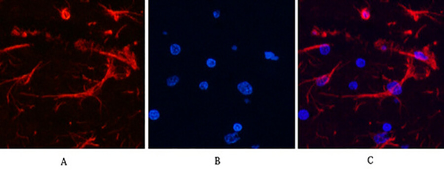
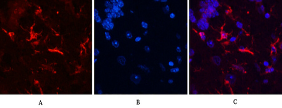
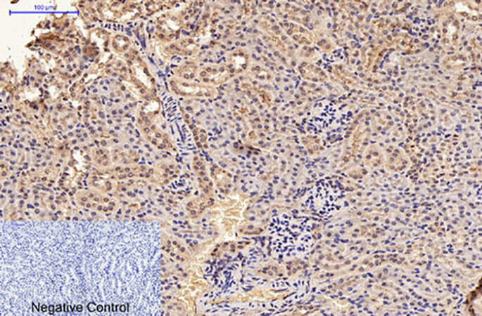
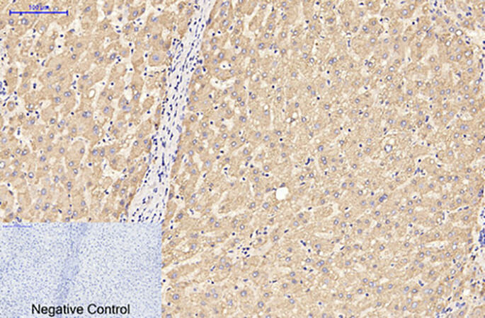
The broad application spectrum of ABM0021 extends across western blot (suggested starting dilutions 1:2000–5000), immunohistochemistry on paraffin-embedded tissue (1:50–300), and immunofluorescence (1:200), with representative images provided on the product page for rat brain, human liver, mouse kidney, rat heart, and mouse brain tissue. The antibody detects endogenous GFAP proteins, and the gene—mapped to chromosomal location 17q21.31—encodes one of the major intermediate filament proteins of mature astrocytes, where it is used as a marker to distinguish astrocytes from other glial cells during development. Mutations in GFAP cause Alexander disease, a rare and typically fatal leukoencephalopathy characterized by the presence of GFAP aggregates in astrocyte cytoplasm, loss of myelin, developmental delay, failure to thrive, and intellectual and motor impairment. The clinical and translational significance of GFAP extends well beyond Alexander disease: a 2024 study validated GFAP as a non-invasive plasma biomarker for glioblastoma and other brain tumors, with high plasma GFAP concentration associated with GBM and lower levels associated with meningiomas and lower-grade gliomas. A 2024 quantum dot-based ratiometric immunoassay for GFAP confirmed that the protein is highly responsive to both ischemic stroke and glioblastoma multiforme, with levels correlating to the extent of brain damage.
The broader competitive landscape for GFAP antibodies has been characterized by the limitations that ABM0021 was engineered to resolve. The Abbkine blog states the comparison directly: traditional GFAP antibodies plague researchers with 30% cross-reactivity against vimentin and nestin, 20% batch-to-batch coefficient of variation, and high backgrounds that obscure low-abundance signals in FFPE brain tissues. These flaws derail publication timelines and waste irreplaceable clinical specimens such as postmortem Alzheimer's disease brains. The epitope masking that occurs during formalin fixation and paraffin embedding is particularly problematic for GFAP, because the intermediate filament assembly buries many epitopes within the coiled-coil polymer core, and antibodies that recognize those buried epitopes produce weak or absent staining in FFPE sections regardless of how much antigen retrieval is performed. ABM0021's epitope in the central rod domain remains accessible after standard citrate-based HIER, a property that the blog attributes to deliberate epitope selection and retrieval optimization.
The antibody formulation reflects competent manufacturing practice. Supplied as a liquid solution at 1 mg/mL in PBS, pH 7.4, containing 0.5% BSA as carrier protein, 0.02% sodium azide as preservative, and 50% glycerol as cryoprotectant, ABM0021 remains stable for one year at -20°C from the date of shipment. The 50% glycerol depresses the freezing point, preventing ice-crystal damage to immunoglobulin protein during storage. Centrifugation of the original vial after thawing and prior to cap removal is recommended for maximum product recovery, and aliquoting is advised to avoid repeated freeze-thaw cycles. Shipping occurs on gel packs with blue ice. The product is for research use only and is not intended for diagnostic or therapeutic applications.
The broader technological context is shifting in directions that will increase demand for epitope-defined, cross-reactivity-free GFAP antibodies. A 2020 milestone review titled "GFAP at 50" noted that glial fibrillary acidic protein arose early in vertebrate evolution, coinciding with the development of different forms of glial cells in the central nervous systems of primitive fish, and that the protein's self-assembly into 10-nm filaments has made it particularly valuable for elucidating the sequences essential for intermediate filament assembly. Spatial proteomics and AI-driven neuro-drug discovery are advancing rapidly, and the Abbkine blog notes that the company is developing a fluorophore-conjugated variant (ABM0021-FITC) for multiplex glial profiling and a knockout-validated rabbit monoclonal for single-cell astrocyte atlas construction. The emerging use of GFAP as a biomarker for astronaut neuroinflammation monitoring in space biology further broadens the application landscape.
What distinguishes ABM0021 from the crowded field of GFAP antibodies is not a single headline specification but the combination of monoclonal epitope definition, zero cross-reactivity to other intermediate filament proteins, a 0.1 ng/mL western blot detection limit, below 3% inter-assay CV, validated performance across western blot, IHC-P, and immunofluorescence in human, mouse, and rat tissue, and independent validation in studies published in Acta Neuropathologica and Journal of Neuroscience. An antibody that generates a clean 45-kDa band on brain homogenate western blots, stains plaque-adjacent astrocytes in Alzheimer's tissue with crisp filamentous resolution, produces clear cytoplasmic signal in primary cortical astrocyte ICC without the 30% background that plagues competitor products, and performs with 99% reproducibility in 1,000-sample-per-week drug screening cascades—that antibody is catalog number ABM0021, priced for the academic laboratory and validated for the translational researcher. The astrocyte marker that refused to blur is now available.
Explore specifications, view representative images, and place your order here: https://www.abbkine.com/product/gfap-monoclonal-antibody-abm0021/


