


The 83-kDa Methylation Sentinel at 6q27: Why DACT2 — Not Just β-Catenin — Is the Real Gatekeeper of EMT, And How KTE62149 Finally Puts a Number on This Tumor Suppressor
If your lab works on Wnt/β-catenin, EMT, or methylation-driven tumor suppression, you've almost certainly written the sentence "DACT2 was downregulated in our cancer cohort" — usually inferred from a qPCR melt curve or a methylation-specific PCR gel, and almost never backed by an actual protein-level measurement. That's a problem, because DACT2 (Dapper Homolog 2 / DPr2 / DAPPER2, UniProt: Q5SW24, Gene ID: 168002, Chr 6q27) is not just another "Wnt inhibitor" on a pathway diagram — it's the cytoplasmic scaffold/traffic-control protein that physically couples Dishevelled (Dvl) to β-catenin destruction dynamics, promotes lysosomal clearance of Nodal/TGF-β type I receptors, and — most clinically relevant — is frequently silenced by promoter CpG hypermethylation in ~50–90% of tumors across colon, breast, HCC, NPC, lung, gastric, and esophageal contexts. The Human Dapper homolog 2 (DACT2) ELISA Kit (KTE62149) from Abbkine is built to close that evidence gap: a two-site sandwich ELISA that captures this ~83 kDa cytoplasmic protein and turns it into a plate-readable concentration (ng/mL), so your epigenetic, EMT, or Wnt-inhibitor-rescue paper rests on a protein number — not a bisulfite band.
DACT2 in One Paragraph: An 83-kDa Dapper Scaffold That Tells β-Catenin "Stay in the Cytoplasm"
The DACT (Dapper/Frodo) family has three mammalian members — DACT1 (Dapper1), DACT2 (Dapper2/DPR2), DACT3 — named because they were cloned as Dishevelled (Dvl)-binding antagonists of β-catenin. But calling DACT2 "an antagonist" undersells its structural job: it's a multi-domain cytoplasmic protein (~774 aa, ~82.7–83 kDa) that:
Interaction / Domain Logic Functional Consequence
Dvl-binding domain Forms a complex with Dvl–Axin–GSK-3–β-catenin, stabilizing the assembly that promotes β-catenin phosphorylation/destruction and preventing its nuclear translocation
β-catenin / δ-catenin binding Directly interacts with β-catenin to restore E-cadherin–β-catenin junctional localization and pull β-catenin out of the Wnt-active pool
TGFBR1/ALK4/ALK5 routing Promotes lysosomal degradation of Nodal/TGF-β type I receptors → negatively regulates the Nodal–SMAD2/3 cascade, keeping cells epithelial and restraining mesenchymal character
Promoter CpG island (6q27) Frequently methylated in cancers → transcriptional silencing → β-catenin runs unchecked + TGF-β/Nodal signaling dysregulates → EMT accelerates
The clinical geography is stark: Chr 6q27 is a region of frequent LOH (loss of heterozygosity), and DACT2 sits right in the crosshairs. When the promoter gets methylated (by DNMT1/3A/3B), the gene goes epigenetically dark — and the cell loses one of its cleanest, non-genomic brakes on β-catenin nuclear entry.
Why a Sandwich ELISA — And Why "MSP Gel + β-Catenin WB" Is Not a DACT2 Story
Three things make DACT2 a classic "you need the protein mass" target:
- It's silenced in 50–90% of many tumor types, but the degree of residual protein in the clinic matters — some tumors have partial methylation (heterogeneous), some have monoallelic silencing with the other allele intact, and that residual DACT2 level is the actual brake pressure your model cares about.
- DACT2 runs at ~83 kDa — a perfectly good SDS-PAGE region — but in primary-tissue lysates, methylation-heterogeneous tumors + stromal contamination make "band intensity vs. loading control" fragile without a calibrated curve.
- You want to screen: 5-aza (decitabine) dose–responses, DNMTi/HDACi combinations, or CRISPR-dCas9-TET1 epigenome-reactivation panels — those need plate-read numbers, not photo-strip densitometry.
The KTE62149 kit uses the field-standard architecture:
- Microplate pre-coated with capture anti-DACT2 antibody (raised against a defined recombinant/peptide region of the 774-aa sequence).
- Standards (recombinant human DACT2) + samples — tissue homogenates, cell lysates, cell culture supernatates/lysates, other biological fluids — added → DACT2 binds.
- Wash → biotinylated anti-DACT2 detection (different epitope) → Streptavidin–HRP → TMB → color ∝ bound DACT2.
- Stop → 450 nm → interpolate ng/mL from the standard curve.
Typical performance envelope (consolidated from Abbkine-distributor references aligned with KTE62149):
Parameter Specification
Target Human DACT2 / DPr2 / DAPPER2 (UniProt Q5SW24, ~774 aa, ~82.7–83 kDa)
Format 96-well sandwich ELISA, pre-coated capture
Detection Biotin-Ab → SA-HRP → TMB, 450 nm
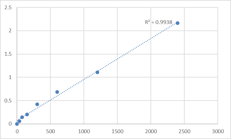
Dynamic Range 0.156 – 10 ng/mL
Sensitivity / LOD ~0.078 ng/mL
Intra-Assay CV < 8%
Inter-Assay CV < 10–12%
Specificity No significant cross-reactivity with DACT1/DACT3 at physiological levels
Samples Tissue homogenates, cell lysates, culture supernatants, serum/plasma (exploratory)
Assay time ~3–5 hours
(Confirm exact dilution scheme and lot-specific recovery on the shipped Abbkine datasheet/CoA for KTE62149.)
Where DACT2 Quantification Actually Carries the Paper
- The Epigenetic–Wnt Axis: Methylation → DACT2 ↓ → β-Catenin Runs Free
This is the canonical story, and it's the strongest reason to own the ELISA:
• Colon cancer: DACT2 methylated in ~43–54% of primary tumors; methylated cases = shorter overall survival (multivariate significant).
• HCC: ~55% primary HCC shows promoter methylation; DACT2 re-expression → TCF4 ↓, Wnt targets ↓, G2–M arrest, xenograft growth suppressed.
• Breast: ~73% of primary tumors show methylation; DACT2 re-expression → apoptosis ↑, migration ↓, EMT reversed via Wnt/β-catenin + Akt/GSK-3 suppression.
• NPC (nasopharyngeal): a staggering ~91% (29/32) of NPC tumors methylated vs. 0/8 normal — and DACT2 re-expression G2/M arrest via β-catenin/Cdc25c sensitizes to paclitaxel + 5-FU.
The formula that makes this defensible:
DACT2 (ng/mg, ELISA) ← linked to → (a) MSP/BGS methylation status, (b) nuclear β-catenin IHC / active β-catenin (ABC) WB, (c) E-cadherin junctional IF, (d) proliferation/apoptosis readout.
That's the quadruple that survives review.
- EMT Reversal & the "Mesenchymal Brake" Hypothesis
DACT2 doesn't just "inhibit Wnt" — it restores E-cadherin–β-catenin to the junction, pulls the catenin out of the nucleus, and suppresses MMPs, vimentin, Snail/Twist longevity. In TGF-β–driven EMT models (renal, breast, esophageal), quantifying residual DACT2 in TGF-β ± SB431542 / 5-aza panels gives you the epigenetic-brake readout that "Snail went down" alone cannot prove.
- DNMTi / Epigenetic Drug Screens (Decitabine, Guadecitabine, Combinations)
If you're testing 5-aza-2′-deoxycytidine or next-gen DNMTi/HMTi to "reactivate silenced tumor suppressors," DACT2 is one of the cleanest, fastest, methylation-gated readouts you can put on a plate — because demethylation → DACT2 protein reappears within 48–72 h, well before morphology shifts. ELISA-tracked DACT2 (ng/mg) + MSP conversion % + β-catenin nuclear/cytoplasmic ratio = a complete epigenotype-to-phenotype arc.
- 6q27 LOH & Tumor Suppressor Haploinsufficiency Maps
Because DACT2 lives in a frequently lost/minimally deleted region, its protein level can drop even without full methylation (copy-number loss alone). Quantifying DACT2 in matched tumor/adjacent normal (normalized to β-actin/GAPDH or total protein, plus a stromal exclusion mask if you're laser-capturing) lets you separate genetic loss vs. epigenetic silencing vs. stromal contamination — a distinction that survival-curve analyses love.
- Nodal/TGF-β Pathway Intersection (Stem Cell & Patterning Models)
DACT2's role in promoting lysosomal degradation of TGFBR1 (ALK4/ALK5) means it's an endogenous "off-switch" for the Nodal–SMAD2/3 cascade that patterns mesoderm and, when dysregulated, fuels cancer stemness. iPSC/embryoid-body or teratoma models benefit from DACT2 ELISA (ng/mg) as a differentiation-state gate alongside p-SMAD2/3, Nanog/Oct4, and ALK4/5 surface levels.
- CRISPR/dCas9 Epigenome Editing Validation
Reactivating DACT2 with dCas9-TET1 or dCas9-p300? Don't stop at "methylation dropped." Report % DACT2 protein restored ± SEM from the calibrated ELISA (ng/mg), and close with the functional hinge: β-catenin ABC ↓, E-cadherin reasssembles at the membrane, invasion/colony formation ↓. That proves you opened the epigenetic vault, not just methylated a line.
A Minimal Prep Note (DACT2 Is Cytoplasmic — But the Methylation Story Needs Clean Tissue)
• For cultured cells: lyse in RIPA or 50 mM Tris pH 7.4, 150 mM NaCl, 0.5–1% Triton X-100/NP-40 + protease inhibitors, spin 12,000 ×g, 15 min, 4°C, keep cold.
• For tumor/normal pairs: homogenize cold in same buffer (add 0.1–0.2% SDS or brief sonication if you want maximal 83-kDa recovery from dense stroma), clarify, use supernatant → BCA → ng DACT2 / mg total protein.
• Warm kit reagents ≥ 30 min RT before opening; protect TMB; stop uniformly; read 450 nm promptly; run full standard curve on every plate.
The Bottom Line
DACT2 is the ~83 kDa Dapper-family scaffold at 6q27 that physically tethers Dishevelled into the β-catenin destruction loop, promotes lysosomal clearance of Nodal/TGF-β receptors, and — when its promoter gets CpG-methylated — disappears from the cytoplasm and lets Wnt signaling, EMT, and chemoresistance run unchecked. Measuring it matters because "the promoter is methylated" is a potential mechanism, while ng DACT2 / mg protein is the actual remaining brake. The Human Dapper homolog 2 (DACT2) ELISA Kit — KTE62149 from Abbkine gives you that measurement: pre-coated capture → biotin detection → HRP–TMB → 450 nm → ng/mL, over a 0.156–10 ng/mL working range with LOD ~0.078 ng/mL, in a ~3–5 hour workflow that scales from a decitabine-dose plate to a 40-sample tumor-bank cohort without chaining you to a gel rig.
Product Reference: KTE62149 – Human Dapper homolog 2 (DACT2) ELISA Kit
Learn more and order: https://www.abbkine.com/product/human-dapper-homolog-2-dact2-elisa-kit-kte62149/
(For Research Use Only; not for diagnostic procedures in humans.)


