


The 227-Amino-Acid Guardian of Protein Time: Why Measuring PCMT1/PIMT — The Cell's L-Isoaspartate Repair Enzyme — Changes How You Read Aging, Neurodegeneration, and Protein Homeostasis
Every protein in your body is slowly, silently, and spontaneously dismantling itself — not by proteolysis, but by a sneaky chemical gremlin called aspartate isomerization and asparagine deamidation. At neutral pH and physiological temperature, the peptide backbone of L-aspartyl (Asp) and L-asparaginyl (Asn) residues cyclizes through an L-succinimide intermediate that, when it opens back up, doesn't always return to normal: ~70–85% of the time it hydrolyzes into L-isoaspartate (L-isoAsp / β-Asp) — a devastating backbone "kink" where a methylene group intrudes into the main chain, destroying the original hydrogen-bonding geometry. These isoaspartate lesions accumulate in long-lived structural proteins (eye-lens crystallins, myelin basic protein, RBC membrane skeletons) and even in signaling proteins (tau, synapsin I, amyloid-β precursors, EIF4EBP2) — and when they do, the protein kinks, aggregates, loses activity, or becomes a template for more damage. Enter PCMT1 — also called PIMT (Protein L-Isoaspartate/D-Aspartate O-Methyltransferase, EC 2.1.1.77) — the cell's only dedicated repair enzyme for this problem: it recognizes the damaged carboxyl group, methylates it using S-adenosylmethionine (AdoMet/SAM) to form a labile methyl ester, which spontaneously decomposes back to the succinimide and then to normal L-aspartate, completing the repair cycle. The Human Protein-L-isoaspartate (PCMT1) ELISA Kit (KTE61292) from Abbkine is built to quantify this unsung repair machine as a calibrated sandwich-ELISA variable (pg/mL) — so your aging, neurodegeneration, or protein-quality-control story rests on a number, not an assumption that "housekeeping enzymes take care of themselves."
PCMT1/PIMT: The Repair Cycle in Plain English
PCMT1 (UniProt: P22061/Q9UBE0, Gene ID: 5110, Chr 6q25.1, 227 aa, computed ~24.8 kDa monomer — runs ~25 kDa on SDS-PAGE) is a highly conserved, cytosolic/partially nuclear S-adenosylmethionine-dependent methyltransferase that initiates the following rescue:
- Spontaneous damage: L-Asp or L-Asn → cyclizes → L-succinimide (Succ) → opens → ~70–85% L-isoAsp (kinked backbone) + ~15–30% normal L-Asp.
- PCMT1 recognition: The enzyme's active site cradles the abnormal isoaspartyl carboxylate.
- Methylation: PCMT1 transfers a methyl group from AdoMet → L-isoAsp-COOH → L-isoAsp-COOCH₃ (methyl ester).
- Spontaneous demethylation: The ester hydrolyzes → reforms the L-succinimide → now has a second chance to reopen as normal L-Asp. Multiple cycles progressively convert the lesion back.
The kicker? Pcmt1-null mice die at ~42 days from a fatal epileptic encephalopathy — massive accumulation of isoAsp-damaged proteins in cortical neurons, continuous EEG abnormality, and catastrophic failure of the brain's most long-lived synaptic and cytoskeletal proteins. Growth retardation in these mice traces back to the seizures, not a global growth-program flaw — a stunning demonstration that "just letting damaged proteins accumulate" has a very short, very violent endpoint.
Why a Sandwich ELISA for PCMT1 — And Why This Isn't "Just a Loading Control"
PCMT1 is soluble, ubiquitous, and ~25 kDa — on paper, an "easy" Western target. But the real research questions are:
• Is PCMT1 protein changing under oxidative stress, aging, alcohol/metabolic liver injury, or neurodegenerative mutation?
• Can we detect it in serum/plasma as a surrogate of tissue repair capacity or extracellular release? (Emerging multi-omics work now links serum PCMT1 to alcoholic liver disease stages and extracellular vesicle/secreted fractions.)
• Do we need to run 40+ samples (time courses, genotypes, cohort tissues) with a CV we can defend?
That's where a two-site sandwich ELISA — pre-coated anti-PCMT1 capture + biotinylated anti-PCMT1 detection → SA-HRP → TMB → 450 nm — beats a gel: you get absolute pg/mL from an on-plate recombinant standard curve, not "band darkness vs. actin."
Assay Principle: KTE61292 Sandwich ELISA
- A microplate is pre-coated with a capture antibody specific for human PCMT1/PIMT.
- Standards (recombinant human PCMT1) and samples — serum, plasma, tissue homogenates, cell lysates, cell culture supernatants/lysates, other biological fluids — are added; PCMT1 present binds.
- After washing → biotinylated anti-PCMT1 detection antibody (different epitope) forms the sandwich.
- Streptavidin–HRP → TMB → color ∝ bound PCMT1.
- Stop → read 450 nm → interpolate unknowns from the PCMT1 standard curve.
Consolidated performance envelope from distributor/technical specs aligned with KTE61292:
Parameter Specification
Target Human PCMT1 / PIMT (UniProt P22061/Q9UBE0, Gene 5110)
Format 96-well sandwich ELISA, pre-coated capture
Detection Biotin-Ab → SA-HRP → TMB, 450 nm
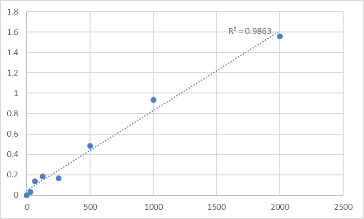
Standard Range 31.2 – 2,000 pg/mL (7-point; equivalently 0.0312 – 2.0 ng/mL)
Sensitivity / LOD ~13.8 – 15.6 pg/mL
Intra-Assay CV < 8–10%
Inter-Assay CV < 10–15%
Specificity Recognizes both natural & recombinant human PCMT1; no significant cross-reactivity with unrelated methyltransferases at physiological levels
Samples Serum, plasma, tissue homogenates, cell lysates, culture supernatants
Assay time ~3–4 hours (per standard protocol)
Status For Research Use Only; not for diagnostic procedures
Where Quantifying PCMT1 Protein Actually Advances the Paper
- Neurodegeneration & the "Aging Protein" Hypothesis
The canonical Pcmt1-KO brain accumulates isoAsp in synapsin I, tau, MAP2, clathrin light chains, α/β-synuclein — the exact substrates whose misfolding drives synaptic failure. In human work, measuring PCMT1 in brain-tissue lysates, CSF (exploratory), or neuron/glia lysates gives you the repair-enzyme reserve that determines whether an isoAsp lesion gets fixed or becomes an aggregate seed. Pair with anti-isoAsp antibodies (e.g., anti-isoD) or isoAsp-targeted LC-MS for the lesion side of the ledger.
- Eye Lens & Crystallin Aggregation / Cataract
This is the most chemically beautiful application: lens fiber cells lose organelles (no proteasome renewal), so their α/β/γ-crystallins live 50+ years and accumulate spontaneous Asp deamidation/isomerization → L-isoAsp → kinked subunits → dissociation of native oligomers → aggregation. PCMT1 is active in the lens epithelium and in fiber-cell remnants; quantifying it in lens homogenates (urea-soluble vs. water-insoluble fractions) and correlating with age/cataract grade is a direct test of the "damage-vs.-repair" balance.
- Alcoholic / Metabolic Liver Disease & "Serum PCMT1"
Recent human data show serum PCMT1 levels decline across alcoholic liver disease stages vs. healthy controls — consistent with a model where chronic hepatocellular stress exhausts repair capacity and/or shifts PCMT1 into vesicle-associated compartments. Running KTE61292 on coded plasma/serum banks lets you test whether PCMT1 adds independent signal beyond ALT/AST/Fib-4.
- Red Blood Cell & Membrane-Protein Longevity (G6PD Deficiency, Sickle, Prosthetic-Valve Shear)
RBC membrane skeletons (band 3/spectrin/anion exchanger complex) accumulate isoAsp under oxidative stress; PCMT1-dependent methylation is the RBC's non-proteasomal "patch" for aged membrane proteins. Measuring PCMT1 in RBC lysates or ghost preparations makes a highly focused quality-control story for hemolysis/hemostasis labs.
- Aging & Long-Lived Protein Turnover Panels
Skin collagen, tendon, myelin basic protein, and vascular ECM proteins all accumulate isoAsp over decades. PCMT1 in biopsy homogenates (normalized to mg total protein, BCA) is the cleanest way to ask: is this tissue still repairing its old proteins, or has it switched to discard-only?
- CRISPR/AAV Validation
Editing PCMT1? Don't just show "mRNA dropped." Report % PCMT1 protein remaining ± SEM from a calibrated curve, normalized to mg total protein, and — if your model allows — link to an isoAsp-readout (anti-isoD IF, methyl-acceptance assay, or MS) so the repair axis is closed.
A Minimal Prep Note (PCMT1 Is Soluble — Treat It Like a Cytosolic Enzyme, Not a Membrane Grind)
• For tissues: homogenize in cold 20–50 mM Tris, pH 7.4, 150 mM NaCl, 0.1–0.5% Triton X-100 or just dounce-homogenize in isotonic buffer + protease inhibitors; clarify ~14,000 ×g, 15 min, 4°C → supernatant is your PCMT1 pool.
• For serum/plasma: process cold/fast; aliquot; avoid >1 freeze–thaw (PCMT1 is stable but low-abundance in circulation, and adsorption losses matter at pg/mL).
• BCA the same lysate → express as pg PCMT1 / mg total protein for tissue or pg/mL for fluids.
• Warm kit reagents ≥ 30 min RT before opening; protect TMB; stop uniformly; read 450 nm promptly; run the full standard curve on every plate.
The Bottom Line
PCMT1/PIMT is the methyltransferase that saves your proteins from their own worst chemistry — the spontaneous Asp/Asn backbone kink that turns a normal residue into a structure-breaking L-isoaspartate and would, left alone, fill neurons with broken synapsins, eye lenses with crystallin aggregates, and membranes with dysfunctional skeletons. Measuring it matters because "it's there" isn't good enough when your experiment turns on whether the cell is repairing or surrendering. The Human Protein-L-isoaspartate (PCMT1) ELISA Kit — KTE61292 from Abbkine gives you the tool to do that: pre-coated capture → biotin detection → HRP–TMB → 450 nm → pg/mL, over a 31.2–2,000 pg/mL standard curve with LOD ~14–16 pg/mL — so the repair axis of your protein homeostasis story has a number you can actually defend.
Product Reference: KTE61292 – Human Protein-L-isoaspartate (PCMT1) ELISA Kit
Learn more and order: https://www.abbkine.com/product/human-protein-l-isoaspartate-pcmt1-elisa-kit-kte61292/
(For Research Use Only; not for diagnostic procedures in humans.)


