


The 20-kDa Gatekeeper of Every Stress Fiber and Cleavage Furrow: Why Total MYL12B Quantification — Not Just Its Phospho-Band — Is the Missing Denominator in Your Contractility Experiment
If your lab works on Rho/ROCK, MLCK, endothelial barrier, cancer invasion, or cytokinesis, you already "measure" the myosin II regulatory light chain every time you run that phospho-specific Western for pSer¹⁹‑MYL12/MRLC2 and call it "pMLC." But here's the uncomfortable question most papers gloss over: you're normalizing a phosphorylation signal to β-actin or GAPDH — two proteins that have nothing to do with the myosin II complex — while ignoring the one variable that actually decides how much phosphorylatable substrate was even there in the first place. The protein in question is MYL12B (aliases MRLC2, MLC-B, MLC20, SHUJUN-1, UniProt: O14950, Gene ID: 103910, Chr 18p11.31, ~172 aa, ~19.7–20.1 kDa computed), the non-muscle myosin II regulatory light chain whose phosphorylation at Ser¹⁹ (by MLCK) and Thr¹⁸/Ser¹⁹ diphosphorylation (by ROCK1/2, MRCK, ZIPK) is the binary switch that turns stress fibers, focal adhesions, cortical tension, and the cytokinetic contractile ring on and off. The Human Myosin regulatory light chain 12B (MYL12B) ELISA Kit (KTE61444) from Abbkine is the tool that finally lets you measure total MYL12B as a calibrated, plate-readable ng/mL — so your pSer¹⁹ signal finally has a biologically meaningful denominator.
MYL12B in One Paragraph: The 172-AA Switch That Decides Whether Your Cell Tenses or Relaxes
MYL12B is not a signaling "readout" floating in the medium — it's a structural subunit permanently assembled into the non-muscle myosin II hexamer (MYH9/MYH10 heavy chains + MYL12A or MYL12B light chains + essential light chain MLC1/3) via its N-terminal EF-hand/Ca²⁺-binding fold and IQ-motif contacts. What makes it special is that the regulatory information lives on MYL12B itself:
• Ser¹⁹ monophosphorylation (MLCK) → 2–3× MgATPase boost, stress-fiber formation competent
• Thr¹⁸+Ser¹⁹ diphosphorylation (ROCK/MRCK/ZIPK) → maximal ATPase (~5×) + robust myosin II filament assembly → cortical tension spikes, adhesion reinforcement, cleavage-furrow constriction
• Dephosphorylation (by MYPT1-PP1cδ complexes and CPI-17 modulation) → myosin II disassembles/fatigue → cortical relaxation
This is why pMLC (pSer¹⁹‑MYL12B) ÷ total MYL12B is the mechanistically honest ratio — and why total MYL12B quantification matters in every Rho/MLCK/ROCK paper that claims "contractility changed."
MYL12B vs. MYL12A: The Paralog Split That Explains "Why Tissue Specificity Happens"
The human genome has two closely related non-muscle MRLC genes:
Gene Alias Size Dominant Expression Quick Rule
MYL12B MRLC2, MLC-B, MLC20 ~172 aa, ~20 kDa Broad: endothelium, epithelium, fibroblasts, platelets, neurons, most non-muscle cells Your "generic non-muscle pMLC" Western is mostly MYL12B
MYL12A MRLC1, MLC-A, smMLC ~171 aa, ~20 kDa Smooth muscle–enriched (aortic, bladder, GI) but also present in non-muscle Smooth muscle contractility panels often track both
Because they're ~85–90% identical in sequence, many "anti-MRLC/pMLC" antibodies recognize both — which is fine for a gel but reinforces why a MYL12B-specific sandwich ELISA (two distinct anti-MYL12B epitopes) is the clean way to lock the ID: you're measuring the non-muscle isoform that runs your endothelial barrier and cancer invasion, not accidentally blending in smooth-muscle contributions.
Why a Sandwich ELISA for a ~20 kDa Light Chain — And Why "Just Blot pSer¹⁹ and Divide by Actin" Is Sloppy
Three structural realities make MYL12B a perfect ELISA target and a problematic gel-only target:
- It's small and runs near the dye front (~20 kDa) — where transfer efficiency, gel-crosslinker crowding, and front-smear artifacts hit hardest. A pre-coated sandwich with two epitopes bypasses all of that.
- It's stoichiometrically limited by the MYH9 hexamer — there's only so much MYL12B per cell, and its levels can shift under chronic Rho/MLCK stimulation or during differentiation (smooth-muscle-lineage transitions). That makes total MYL12B pool size a variable, not a constant.
- Your pSer¹⁹ signal is meaningless without the substrate pool. If MYL12B drops 30% because of isoform switching or chronic ROCK inhibition, "pMLC band looks fainter" could mean "less kinase activity" or "less light chain to phosphorylate" — and the wrong conclusion changes the paper.
The KTE61444 kit uses the proven architecture:
- Microplate pre-coated with anti-MYL12B capture antibody.
- Standards (recombinant MYL12B) + samples — serum, plasma, tissue homogenates, cell lysates, culture supernatants — added → MYL12B binds.
- Wash → biotinylated anti-MYL12B detection antibody (different epitope) → Streptavidin–HRP → TMB → color ∝ bound MYL12B.
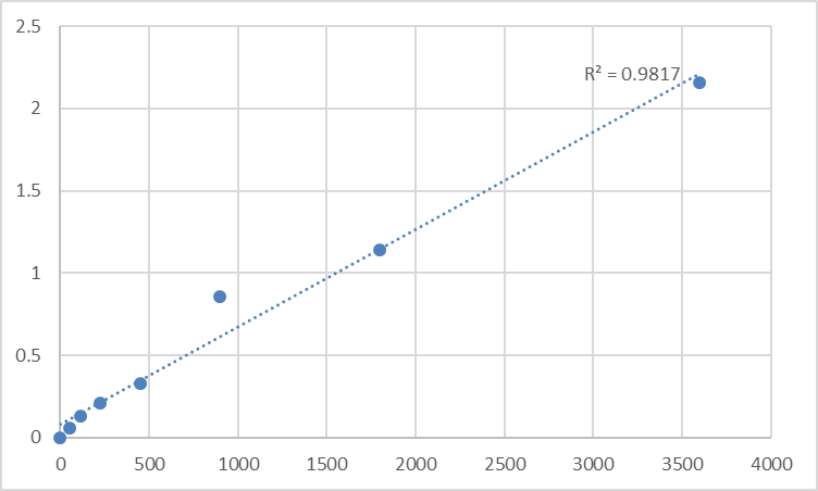
- Stop → 450 nm → interpolate MYL12B concentration from the standard curve.
Consolidated performance envelope (matches distributor consensus across this kit family):
Parameter Specification
Target Human MYL12B / MRLC2 (UniProt O14950, Gene 103910)
Format 96-well sandwich ELISA, pre-coated capture
Detection Biotin-Ab → SA-HRP → TMB, 450 nm
Dynamic Range 0.312 – 20 ng/mL
Sensitivity / LOD ~0.114 ng/mL
Intra-Assay CV < 8–10%
Inter-Assay CV < 10–12%
Samples Tissue homogenates, cell lysates, cell culture supernatants, serum, plasma, other biological fluids
Assay time ~3–4 h
(Confirm exact dilutions and lot-specific recovery on the shipped Abbkine datasheet for KTE61444.)
Where Total MYL12B Quantification Actually Fixes the Story
- Endothelial Barrier & Permeability (VE-cadherin / Rho/ROCK axis)
Thrombin, histamine, LPS, or shear stress → ROCK → MYPT1 pThr⁶⁹⁶/pThr⁸⁵³ → MLC diphosphorylation → AJ contraction → permeability ↑.
The gold-standard readout should be: pSer¹⁹-MYL12B (WB) ÷ total MYL12B (ELISA, ng/mg protein) — not pMLC/actin. TEER dropouts, dextran-FITC leakage assays, and VE-cadherin internalization all become interpretable when the phosphorylation ratio is honest.
- Cancer Invasion, EMT & the Contractile-Mesenchymal Transition
Invasive cells need front-to-back cortical tension (ROCK/pMLC high at the rear, Rac/WAVE at the leading edge). Chronic ROCK inhibition or MYL12B knockdown collapses stress fibers and focal adhesions but also (counterintuitively) can enhance amoeboid-to-mesenchymal plasticity in some contexts. Quantifying total MYL12B across sgRNA conditions, dose–response (Y-27632/fasudil), or EMT inducers (TGF-β) gives you the substrate pool that explains whether pSer¹⁹ shifted because of kinase or because the light chain pool changed.
- Cytokinesis & Cleavage-Furrow Mechanics
The contractile ring is a MYH9–MYL12B–Anillin–RhoA–ECT2 structure. During anaphase, diphosphorylated MYL12B concentrates at the equator and drives furrow ingression (~2–5 µm/min in HeLa). If your experiment tests ECT2 depletion, Rock inhibitor washout, or Aurora B kinetics, the control lane should include total MYL12B (ELISA, pg/µg protein) so "pSer¹⁹ dropped" isn't misinterpreted as a pure kinase effect.
- Smooth Muscle & Vascular Tone (Where MYL12A/MYL12B Ratio Also Matters)
Aortic rings, pulmonary artery, bladder strips — MYL12A dominates but MYL12B is present, and some non-smooth-muscle stromal/fibroblast contamination rides on the MYL12B pool. A specific ELISA helps you partition "smooth-muscle MRLC" vs. "infiltrate/fibroblast MRLC" when you're dissecting KCl vs. agonist (ET-1, AngII, UTP) vs. ROCK contributions.
- Platelet Activation & Thrombosis
Platelets are packed with MYL12B (non-muscle myosin IIA = MYH9/MYL12B), and its phosphorylation drives shape change, granule secretion, and clot retraction. Measuring total MYL12B in platelet lysates (normalized to protein or MYH9) as a baseline for pSer¹⁹ dynamics is a cleaner PD readout in anti-platelet drug screens.
- CRISPR/siRNA Validation
Editing MYL12B? Report % MYL12B protein remaining ± SEM from the calibrated curve (ng/mg, BCA), not just "pMLC band / actin." And if you knocked it out successfully, show that pSer¹⁹ signal collapses because the substrate vanished — that's the mechanistic rigor that gets picked up in review.
A Minimal Prep / Normalization Rule So Your ng/mL Means Something
• Lysates: RIPA or 50 mM Tris pH 7.4, 150 mM NaCl, 0.5–1% NP-40 + protease + phosphatase inhibitors + 1 mM Na₃VO₄ + 10 mM NaF (keep MYL12B phosphorylation state intact if you're running the parallel pSer¹⁹ Western). Clarify 12,000–16,000 ×g, 15 min, 4°C.
• Tissue: homogenize cold in the same buffer; spin; keep supernatant.
• BCA the same lysate → express as ng MYL12B / mg total protein.
• Warm kit reagents ≥ 30 min RT before opening; protect TMB; stop uniformly; read 450 nm promptly; run the full standard curve on every plate.
The Bottom Line
MYL12B is the 172-amino-acid, 20-kDa regulatory light chain that sits at the exact hinge where Rho/ROCK/MLCK signaling becomes force: it's the substrate whose phosphorylation state is the actomyosin switch for every stress fiber, focal adhesion, and cleavage furrow in your non-muscle cells. Measuring it as a calibrated variable — not assuming it's constant, not dividing pSer¹⁹ by actin — is what separates a real contractility analysis from a gel-based suggestion. The Human Myosin regulatory light chain 12B (MYL12B) ELISA Kit — KTE61444 from Abbkine gives you that variable: pre-coated capture → biotin detection → HRP–TMB → 450 nm → ng/mL, over a 0.312–20 ng/mL range with LOD ~0.114 ng/mL, in a ~3–4 hour workflow that slots next to your pSer¹⁹ Western and TEER/scratch assays as the denominator the field forgot to measure.
Product Reference: KTE61444 – Human Myosin regulatory light chain 12B (MYL12B) ELISA Kit
Learn more and order: https://www.abbkine.com/product/human-myosin-regulatory-light-chain-12b-myl12b-elisa-kit-kte61444/
(For Research Use Only; not for diagnostic procedures in humans.)


