


The Only Motor That Walks Backward: Why Myosin-VI (MYO6) Is the Secret Metastasis Driver Hiding in Your Endocytic Pit — And How KTE61414 Puts a Number on It
There are 14 classes of myosin motors in humans, and almost every introductory cell-biology lecture picks the same poster children: Myosin-II for contractility, Myosin-V for long-range cargo kinesin-like runs, and Myosin-I for membrane tension. But the molecule that quietly runs the most counterintuitive — and arguably the most consequential — transport logic in the mammalian cell is Myosin-VI (MYO6), the only known minus-end-directed actin-based motor, the only myosin whose lever-arm insertion flips its polarity, and the cargo-handling workhorse that decides whether a nascent clathrin pit actually pinches off, whether your stereocilia stay stiff or collapse into deafness, and whether a cancer cell can polarize endocytic traffic toward invasive membrane ruffles. The Human Myosin-VI (MYO6) ELISA Kit (KTE61414) from Abbkine is built to give you a calibrated, two-site sandwich ELISA readout of this ~182 kDa heavy chain — so your endocytosis, Golgi-trafficking, hearing-biology, or invasion story rests on ng/mL interpolated from a recombinant standard curve, not a "smear at 180 kDa that might be MYO6 if the antibody didn't cross-react with MYO5."
MYO6 in One Paragraph: The Reverse Gear of the Actin Highway
MYO6 (UniProt: Q00499, Gene ID: 4646, Chr 6q14.1, computed ~182.7 kDa heavy chain / ~1513 aa before alternative splice) is a dimer/tetramer-forming motor with a canonical N-terminal motor domain (ATP- and actin-binding, with the familiar P-loop Walker A/B switch), a long coiled-coil stalk (3 IQ calmodulin/light-chain binding motifs), and a C-terminal globular tail domain (CTD / cargo-binding region) that engages a surprisingly diverse adaptor network:
Adaptor / Binding Partner Functional Context
DAB2 (Disabled-2) / TOM1 / TOLLIP Clathrin-mediated endocytosis (CME) pinch-off & uncoating — MYO6 anchors the vesicle to actin to prevent retrograde sliding
GIPC / synectin (+ RUFY3) Receptor trafficking (GFRs, integrins, ion channels) — polarizes cargo delivery to specialized membrane domains
OPTN (Optineurin) Links MYO6 to autophagy (selective autophagic receptor crosstalk) and immune signaling; OPTN mutations in ALS/glaucoma intersect this node
GOLGA2 / GM130 Golgi-to-plasma-membrane / secretory vesicle routing
SAP97 / hDlg / NHERF1 Polarized epithelial transport (proximal tubule, intestine, cochlear hair cells)
The mechanical headline: every other myosin walks toward the barbed (+) end (cell periphery). The small 3-α-helix insert in MYO6's converter domain torques the lever arm so the power stroke points the opposite way — toward the pointed (-) end, i.e., inward from the plasma membrane. That single geometric inversion is why MYO6 can pull freshly budded vesicles away from the membrane into the cortical actin forest instead of letting Brownian motion drift them back into the surface.
Why a Sandwich ELISA for a ~183 kDa Motor — And Why "Just IP/WB It" Leaves Money on the Table
MYO6 is large, filament-forming, and cargo-adaptor-associated, which creates three practical headaches for gel-only quantification:
- The ~180 kDa zone is messy — especially in brain, kidney, and epithelial lysates where other myosins (MYH9/10 non-muscle MHC at ~220 kDa, MYO5A/B at ~210 kDa) run nearby, and where cross-reactive heavy-chain antibodies can haunt you. A two-epitope sandwich (pre-coated capture + biotin detection, different tail/motor-region epitopes) is your insurance.
- MYO6 levels shift subtly in cancer and hearing loss, not 10-fold like a cytokine — so the experiment needs pg–ng sensitivity and CVs, not a "band vs. actin" eyeball.
- You want to run panels: siRNA dose responses, DAB2/MYC/Rac1 invasiveness grids, cochlea-organoid harvests, or clinical FFPE-lysate banks — and that scales as plates, not gels.
The KTE61414 format is the classic Abbkine pre-coated sandwich ELISA:
- Microplate pre-coated with anti-MYO6 capture antibody.
- Standards (recombinant human MYO6 / defined calibrator) + samples — tissue homogenates, cell lysates, cell culture supernatants/lysates, other biological fluids — added → MYO6 binds.
- Wash → biotinylated anti-MYO6 detection antibody (different epitope) → Streptavidin–HRP.
- TMB → stop → 450 nm → interpolate MYO6 concentration from the standard curve.
Typical performance envelope for this kit class (confirm exact range/recovery on your lot's CoA):
Parameter Typical KTE61414-class spec
Target Human MYO6 / Myosin-VI heavy chain (UniProt Q00499, ~182 kDa)
Format 96-well sandwich ELISA, pre-coated capture
Detection Biotin-Ab → SA-HRP → TMB, 450 nm
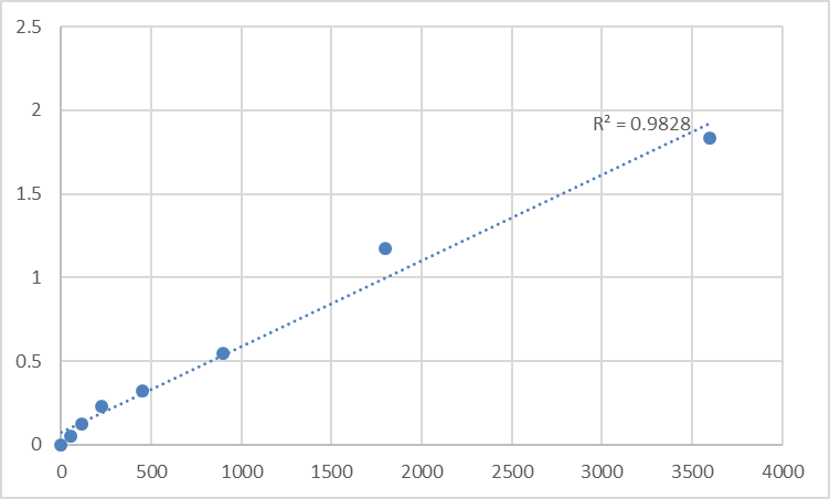
Dynamic Range 0.156 – 10 ng/mL (7-point standard)
Sensitivity / LOD ~0.05–0.09 ng/mL
Intra-Assay CV < 7–8%
Inter-Assay CV < 10%
Samples Tissue homogenates, cell lysates, culture supernatants, other biological fluids
Assay time ~3–5 hours
(Lock your Methods citation to the shipped Abbkine datasheet for KTE61414.)
Where Quantifying MYO6 Actually Carries the Paper
- Cancer Invasion & the "Endocytic Polarity" Model
This is the fastest-growing MYO6 story. Multiple independent screens have flagged MYO6 as recurrently amplified / overexpressed in ovarian, breast, colorectal, and pancreatic cancers, where it drives invasion by polarizing clathrin-mediated endocytosis and integrin-trafficking toward leading-edge ruffles, and by coupling to MT1-MMP delivery and invadopodia turnover. The mechanistic shorthand: more MYO6 → more directional cargo pull → more invasive membrane dynamics.
Report it as ng MYO6 / mg total protein (BCA) across siMYO6 / shMYO6 / CRISPR lines, paired with Matrigel invasion, DAB2/integrin β1 recycling, and actin-rich ruffle IF — that's the triple that survives review.
- Hereditary & Acquired Hearing Loss (Stereocilia Maintenance)
MYO6 is the poster child for actin-motor roles in inner-ear hair-cell stereocilia: it anchors the tip-link / ankle-link complex and pulls opposing actin filaments to maintain tension in the bundle. Mutations cause:
• DFNA22 — autosomal dominant progressive hearing loss (missense in motor/tail)
• DFNB37 — autosomal recessive profound congenital deafness (null/frameshift)
Quantifying MYO6 protein in cochlear microdissections, organ of Corti explants, or (in preclinical models) vestibular/lateral-wall lysates gives you a direct readout of whether a toxin, noise protocol, or rescue transgene actually restored motor levels — not just "the ABR threshold shifted."
- Clathrin-Mediated Endocytosis (CME) Flux & Kidney/Gut Epithelial Polarity
In proximal tubule epithelial cells (PTECs) and intestinal enterocytes, MYO6–DAB2/NHERF1 complexes organize the apical endocytic coat and vesicle scission. Knockdown produces clathrin pit accumulation, defective receptor (Megalin/Cubilin/NaPi-IIa) turnover, and polarity stress. An ELISA lets you run siRNA dose–responses and correlate MYO6 levels with transferrin uptake, dextran-TR, or receptor half-life without camping on a TEM.
- Autophagy & Selective Autophagic Cargo Routing (OPTN/TOM1 Axis)
MYO6 interacts with OPTN (ALS/glaucoma-linked) and TOM1 (IL-1R/Tollip ESCRT-0 routing) to position autophagic precursors near the actin cortex and modulate TLR/cytokine receptor turnover. In neurodegeneration and neuroinflammation models (BV2/primary microglia, iPSC-neurons), quantifying MYO6 alongside LC3-II/LC3-I, p62/SQSTM1, and OPTN frames the motor as a trafficking gate, not just a transport footnote.
- CRISPR/AAV / Rescue Validation
Editing MYO6? Don't stop at "band fainter." Give reviewers:
• % MYO6 protein remaining ± SEM from the calibrated ELISA (ng/mg)
• Confirmatory IF showing loss from stereocilia-like apical fringes or cortical puncta
• Functional payout: invasion index, ABR threshold, transferrin uptake, or LC3 flux — whatever fits your model
That combination is what elevates a MYO6 paper from "we knocked it out" to "we characterized the motor node."
A Minimal Prep Blueprint (MYO6 Is a Large, Actin-Adjacent Motor — Treat the Cortex Right)
• For cultured cells: lyse in RIPA or 0.5–1% NP-40 / 50 mM Tris pH 7.4 / 150 mM NaCl + protease inhibitors + 1–2 mM ATP (optional, stabilizes motor conformations during prep), keep cold, clarify 12,000–16,000 ×g, 15 min, 4°C.
• For tissue (cochlea, kidney cortex, tumor): homogenize cold in the same buffer + 0.1% SDS or 0.5% deoxycholate optional if membrane cargo complexes resist gentle detergent; spin hard; keep supernatant.
• BCA the same final lysate → express as ng MYO6 / mg total protein.
• Warm kit reagents ≥ 30 min RT before opening; protect TMB from light; stop uniformly; read 450 nm promptly; and — critical — run the full standard curve on every plate (large motor proteins can adsorb subtly to plastic; the curve is your calibration anchor).
The Bottom Line
MYO6 is the 182 kDa reverse-direction actin motor that pulls vesicles inward when every other myosin pushes outward — and that geometrical flip is exactly why it controls clathrin pinch-off, stereocilia tension, polarized epithelial cargo, and the endocytic polarity that cancer cells hijack to invade. Measuring it deserves more than a "180 kDa band vs. GAPDH" photo. The Human Myosin-VI (MYO6) ELISA Kit — KTE61414 from Abbkine gives you the right architecture: pre-coated capture → biotin detection → HRP–TMB → 450 nm → ng/mL, over a 0.156–10 ng/mL working range, in a ~3–5 hour workflow that scales across genotypes, treatments, and tissue cohorts without chaining you to a transfer stack.
Product Reference: KTE61414 – Human Myosin-VI (MYO6) ELISA Kit
Learn more and order: https://www.abbkine.com/product/human-myosin-vi-myo6-elisa-kit-kte61414/
(For Research Use Only; not for diagnostic procedures in humans.)


