


The Scaffold That Tames the HECT E3: Why Quantifying N4BP1 Changes How You Read Ubiquitin Signaling, Selective Autophagy, and Innate Immune Crosstalk
Everyone obsessed with ubiquitin signaling talks about the E3 ligases — SKP1–CUL1–F‑box, APC/C, and the HECT clan — but the molecule that often decides whether the ligase actually gets to act is a non-enzymatic, multi-domain scaffold that refuses to fit neatly into a single pathway name. That molecule is N4BP1 (NEDD4-binding protein 1, aliases NEDD4L interactor / KIAA0619-like, UniProt: Q86UW9, Gene ID: 55842) — a ~130–140 kDa zinc-finger/RING-like and coiled-coil–rich protein that binds the NEDD4/NEDD4L (NEDD4-1/NEDD4-2) HECT E3 ubiquitin ligases, interfaces with selective autophagy receptors, and has emerged as a critical node in TNFR/NF-κB regulation, IFN responses, and the suppression of aberrant RIPK1-dependent cell death (necroptosis/apoptosis) under genotoxic or inflammatory stress. The Human NEDD4-binding protein 1 (N4BP1) ELISA Kit (KTE61399) from Abbkine is built to give you a calibrated, two-site sandwich ELISA readout of total N4BP1 protein — so you stop treating this pivotal scaffold like "the 130 kDa smear that co-immunoprecipitates with NEDD4" and start treating it as a quantifiable variable you can plot across genotypes, treatments, and patient-derived lysates.
N4BP1 in One Paragraph: A Scaffold, Not a Ligase, That Still Controls Who Lives and Who Dies
Unlike most ubiquitin-focused papers, N4BP1 has no catalytic ubiquitin ligase or protease domain of its own. Its power is architectural:
• NEDD4/NEDD4L interaction surface → positions or dampens the HECT E3 relative to substrates (traffic, endocytosis, membrane protein turnover — think ENaC, Notch, RTKs, integrins, TGF-β receptors)
• RIPK1 / TNFR1 complex crosstalk → recent work frames N4BP1 as a negative regulator of RIPK1 kinase activity and a stabilizer of the TNFR1–complex I survival signal, so that under FADD/CASP8 suppression the cell doesn't slide catastrophically into necroptosis — loss or disruption of N4BP1 axis tilts the balance toward RIPK1-dependent cell death
• Selective autophagy & pathogen-host interfaces → N4BP1 can associate with autophagy adaptors (e.g., via NBR1/SQSTM1-related crosstalk in specific contexts) and host-defense circuits, placing it in the same conceptual neighborhood as xenophagy and IFN-stimulated turnover
• Cancer & metabolic stress → N4BP1 levels modulate how epithelial and immune cells handle stress-induced ubiquitin flux; in some cancers, its expression correlates with the ability to restrain excess RIPK1-driven inflammation
In short: N4BP1 is a stress-responsive rheostat that couples "how ubiquitin is wired" to "whether the cell chooses apoptosis, necroptosis, or survival signaling."
Why a Sandwich ELISA for N4BP1 — And Why the "130 kDa Band" Is a Weak Claim
Because N4BP1 is large, multi-domain, and often localized to cytosolic puncta / stress granules / membrane-proximal scaffolds, your typical Western suffers from:
- Post-translational heterogeneity (phosphorylation, ubiquitination, SQSTM1-p62 crosstalk) that smears the 130 kDa region and makes "fold change" noisy.
- Epistatic environments: N4BP1 can be recruited into complexes and degraded under specific stress (or stabilized when the E3 is inhibited) — so "band intensity" confounds protein amount with turnover state.
- Throughput: once you move to genotype panels, drug-dose matrices, or tissue-bank lysates, gel-only quantification breaks.
A two-site sandwich ELISA (pre-coated capture + biotinylated detection → SA-HRP → TMB → 450 nm) gives you:
• Two independent anti-N4BP1 epitopes → higher specificity in complex lysates
• A recombinant N4BP1 standard curve on every plate → OD → interpolated ng/mL / pg/mL
• Scalable 96-well format for time courses, CRISPR screens, and cohort normalization
Assay Principle: KTE61399 — Sandwich ELISA (Pre-Coated)
The kit uses the classic architecture you've seen across the Abbkine ELISA family:
- A microplate is pre-coated with a capture antibody specific for human N4BP1.
- Standards (recombinant human N4BP1) and samples — tissue homogenates, cell lysates, cell culture supernatants/lysates, other biological fluids — are added; N4BP1 present binds.
- After washing → biotinylated anti-N4BP1 detection antibody (different epitope) forms the sandwich.
- Streptavidin–HRP → TMB → color ∝ bound N4BP1.
- Stop → read Absorbance at 450 nm → interpolate unknowns from the N4BP1 standard curve.
A representative performance envelope commonly cited for this kit class (always cross-check your shipped CoA):
Parameter Typical KTE61399-class specification
Target Human N4BP1 (NEDD4-binding protein 1, UniProt Q86UW9, Gene ID 55842)
Format 96-well sandwich ELISA, pre-coated capture
Detection Biotin-Ab → SA-HRP → TMB, 450 nm
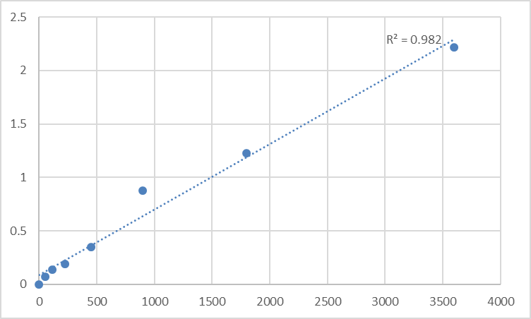
Dynamic Range 0.156 – 10 ng/mL (7-point standard)
Sensitivity / LOD ~0.06–0.10 ng/mL
Intra-Assay CV < 8%
Inter-Assay CV < 10–12%
Samples Tissue homogenates, cell lysates, culture supernatants, other biological fluids
Assay time ~3–5 hours
(Exact range, dilution factors, and recovery should be read from the lot-specific datasheet on the Abbkine product page.)
Where Quantifying N4BP1 Protein Actually Advances the Paper
- TNFR1–RIPK1 Cell-Death Decisions (Necroptosis / PANoptosis Crosstalk)
If your model inhibits CASP8 (zVAD, vFLIP, Caspase-8 conditional KO) or perturbs FADD–RIPK1–complex I stability, the crucial question becomes: does RIPK1 kinase activity run unchecked? N4BP1 is positioned as a braking/stabilizing element in that system. Quantify N4BP1 (normalized to mg total protein, BCA) alongside:
• p-RIPK1 (Ser166/Thr467 reporters)
• p-MLKL (Ser358/Thr357)
• Caspase-8 cleavage, LDH release, PI/annexin V
→ you move from "death happened" to why the rheostat slipped.
- NEDD4/NEDD4L Substrate Traffic & RTK/ENaC Turnover
N4BP1 helps define how the NEDD4 E3 axis handles membrane cargo. In kidney/collecting-duct models (ENaC), neuroblastoma, and receptor-tyrosine-turnover contexts, tracking N4BP1 levels tells you whether the scaffold is present to bias the ligase toward (or away from) a substrate pool — a subtle but publishable regulatory layer beyond "NEDD4 went up."
- Infection, Xenophagy & IFN Responses
N4BP1 sits near the intersection where ubiquitin-coated bacteria are routed toward selective autophagy (xenophagy) and where IFN/TLR signaling meets cell-death governance. In macrophage/ dendritic-cell systems challenged with intracellular bacteria or DNA/RNA mimics, N4BP1 quantification in cell lysates + LC3-II/ p62 flux creates a clean "host-routing" narrative.
- Cancer Cell Stress (Genotoxic, Nutrient, or Proteotoxic)
Some tumors exploit the RIPK1 cell-death threshold and the ubiquitin routing that constrains it; N4BP1 modulation (CRISPR or drug-induced stress) can shift the balance toward inflammatory cell death vs. survival. Report % N4BP1 protein remaining ± SEM from the calibrated curve, normalized to total protein, and tie it to clonogenic survival or orthotopic growth so the scaffold isn't just a "binder" but a phenotype driver.
- CRISPR/AAV Validation
Editing N4BP1? Don't just show a "130 kDa band fainter." Give readers:
• ELISA-derived ng N4BP1 / mg protein (mean ± SEM, n≥3)
• A confirmatory IF/tissue-IFI (if you have it) to show distribution (punctate/cytosolic vs. stress re-localization)
• The functional hinge: RIPK1-dependent death readout or TNFR1 signaling readout (p-IKK, p-p65, NF-κB reporter)
Reviewers consistently reward that triad.
A Minimal Prep Note (N4BP1 Is Large, Cytosolic, and Stress-Granule Friendly)
• For cultured cells: lyse in RIPA or 0.5–1% NP-40 / 50 mM Tris pH 7.4 / 150 mM NaCl + protease inhibitors (+ 1–5 mM NEM if you want to limit deubiquitination during prep); clarify 12,000–16,000 ×g, 15 min, 4°C; keep cold.
• For tissue (spleen, kidney, tumor): homogenize cold in the same buffer; spin; use supernatant.
• BCA the same final lysate → express as ng N4BP1 / mg total protein.
• Warm kit reagents ≥ 30 min RT before opening; protect TMB from light; stop uniformly; read 450 nm promptly; and run the full standard curve on every plate — scaffold proteins with punctate/aggregate-adjacent distributions are exactly where plate-to-plate drift hides.
The Bottom Line
N4BP1 is the ~130 kDa, non-enzymatic scaffold that binds NEDD4-family HECT ligases, helps govern TNFR1–RIPK1 cell-death decisions, and threads ubiquitin routing, selective autophagy, and innate immune homeostasis into a single survival-or-demise calculus. Measuring it as a calibrated ELISA variable instead of a gel-shadow makes your ubiquitin/necrosis paper quantitatively credible. The Human NEDD4-binding protein 1 (N4BP1) ELISA Kit — KTE61399 from Abbkine gives you that architecture: pre-coated capture → biotin detection → HRP–TMB → 450 nm → ng/mL, in a 0.156–10 ng/mL working envelope with LOD in the ~0.06–0.1 ng/mL bracket — so the "binder that controls the ligase" becomes a number you can defend.
Product Reference: KTE61399 – Human NEDD4-binding protein 1 (N4BP1) ELISA Kit
Learn more and order: https://www.abbkine.com/product/human-nedd4-binding-protein-1-n4bp1-elisa-kit-kte61399/
(For Research Use Only; not for diagnostic procedures in humans.)


